C-fos is a proto-oncogene that is the human homolog of the retroviral oncogene v-fos. In the neuroscience field, immuno-labeling of the c-fos protein has been used to study neuronal activity patterns in several vertebrate brain systems. Expression of c-fos is an indirect marker of neuronal activity because c-fos is often expressed when neurons fire action potentials. We have used the technique (see below) to explore frequency coding and spontaneous activity patterns in the auditory brainstem and midbrain.
Tonotopic organization in the auditory midbrain
We have developed a c-fos immunolabeling technique to study the tonotopic organization of the inferior colliculus (IC) in the chinchilla.
FIGURE A shows a band of c-fos labeling in IC after stimulation with a 2 kHz tonal signal. We verified that the neurons in this region were responding best to 2 kHz as shown in the neural frequency tuning curve derived by single unit electrophysiological recording. The histogram plots the c-fos activity across the IC tonotopic axis.
FIGURE B shows the c-fos band after 6 kHz frequency stimulation. Again the right hand histogram shows the amount of labeling across the IC tonotopic array. Note in both figures A and B, the c-fos peak is flanked by regions of much reduced labeling. This could represent lateral inhibitory effects.


Changes in neural activity patterns
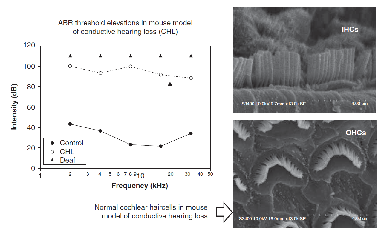
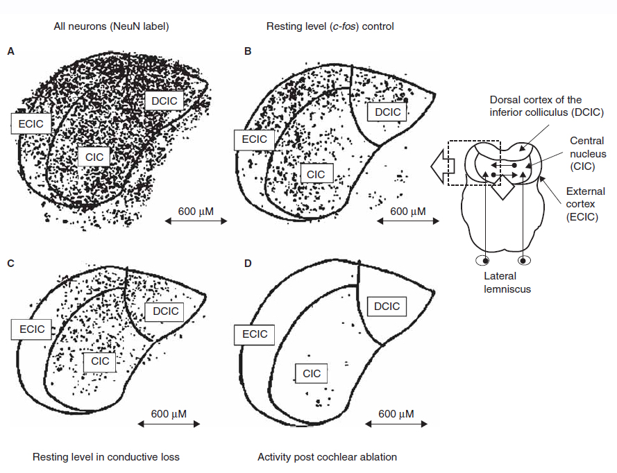
We have also used c-fos immunolabeling to study changes in resting neural activity patterns in the brainstem and midbrain during conductive hearing loss.
The graph on the right shows an increase in hearing thresholds in subjects with conductive hearing loss, although no changes were seen to the inner and outer hair cells in the cochlea of these subjects.
Resting levels of spontaneous neural activity was measured using c-fos as a marker in both the brainstem and the midbrain (inferior colliculus). Control subjects were compared to those with conductive hearing loss (CHL) and those that were deaf.
Results from both the cochlear nucleus and midbrain showed that although CHL does not cause obvious change to the cochlea, it results in significantly decreased resting levels of neural activity through the auditory pathway from the brainstem to the midbrain.
References
D’Alessandro L. and Harrison R.V. Excitatory and inhibitory tonotopic bands in chinchilla inferior colliculus revealed by c-fos immune-labeling. Hearing Research. (2014) 316: 122-128.
Harrison R.V. and Negandhi J. Resting neural activity patterns in auditory brainstem and midbrain in conductive hearing loss. Acta. Oto-Laryngologica. (2012) 132(4): 409-414.