Electron microscopes use a beam of accelerated electrons to image the structure of a wide range of biological and inorganic specimens. In our lab, we have used both scanning and transmission electron microscopy to view specific parts of the inner ear.
Scanning electron microscopy
A scanning electron microscope (SEM) produces high magnification images by using a beam of electrons that interacts with atoms on the surface of the sample. Images produced by the SEM show the topography of the surface of the sample and its resolution can be as low as 1 nanometre. Below are some of the images we have obtained using SEM on various samples of the cochlea.
Click on any photo to obtain a higher resolution image that can be downloaded (copy or save image).
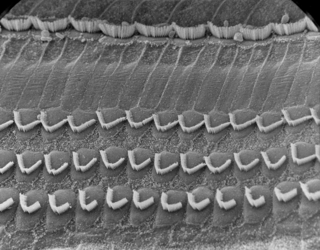
Surface view of three rows of outer hair cells and one row of inner hair cells in the mid-turn of the (chinchilla) cochlea.
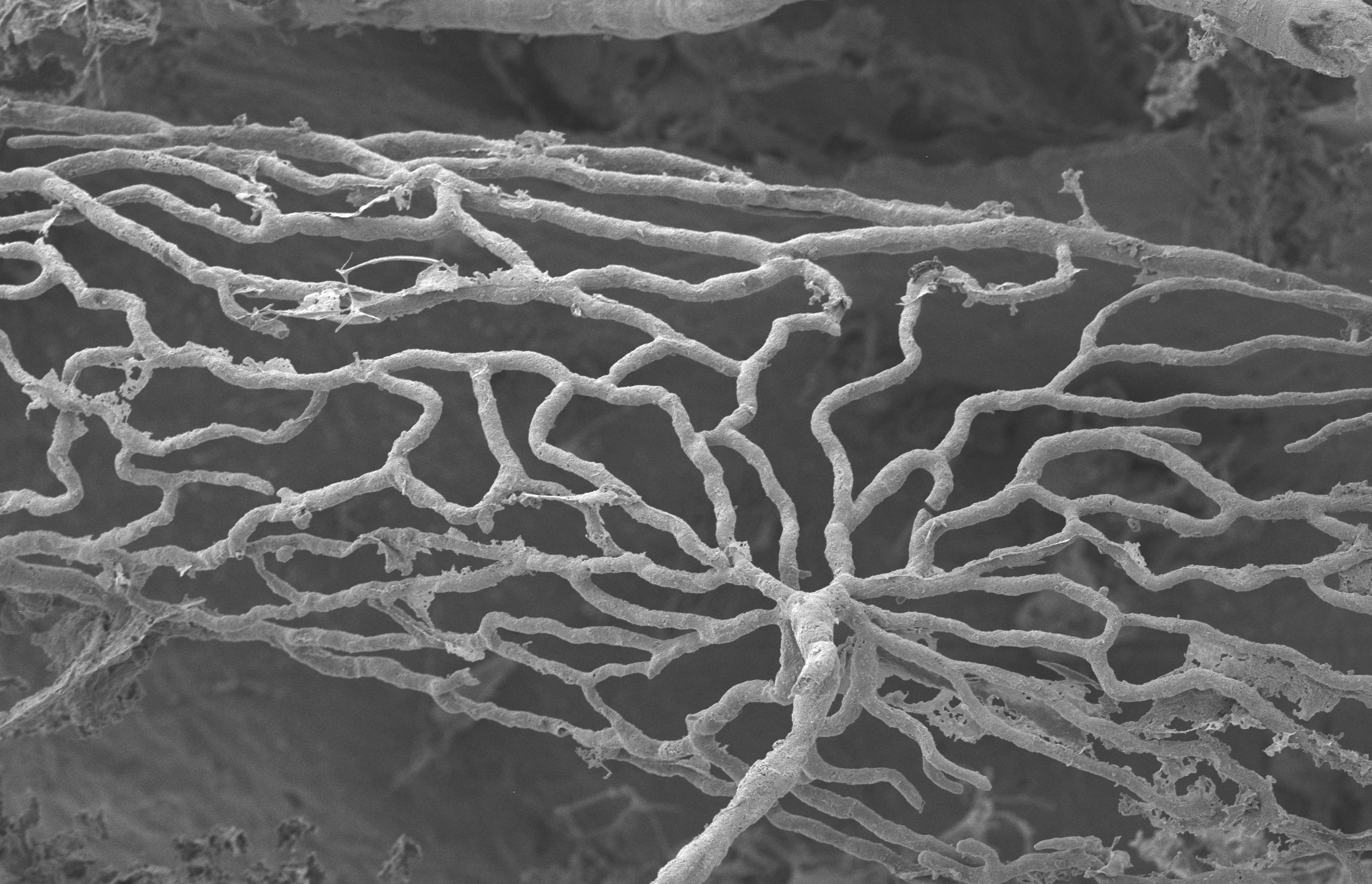
Image of a corrosion cast of the stria vascularis of the mouse cochlea. Note the fan-like structure of the capillary network.
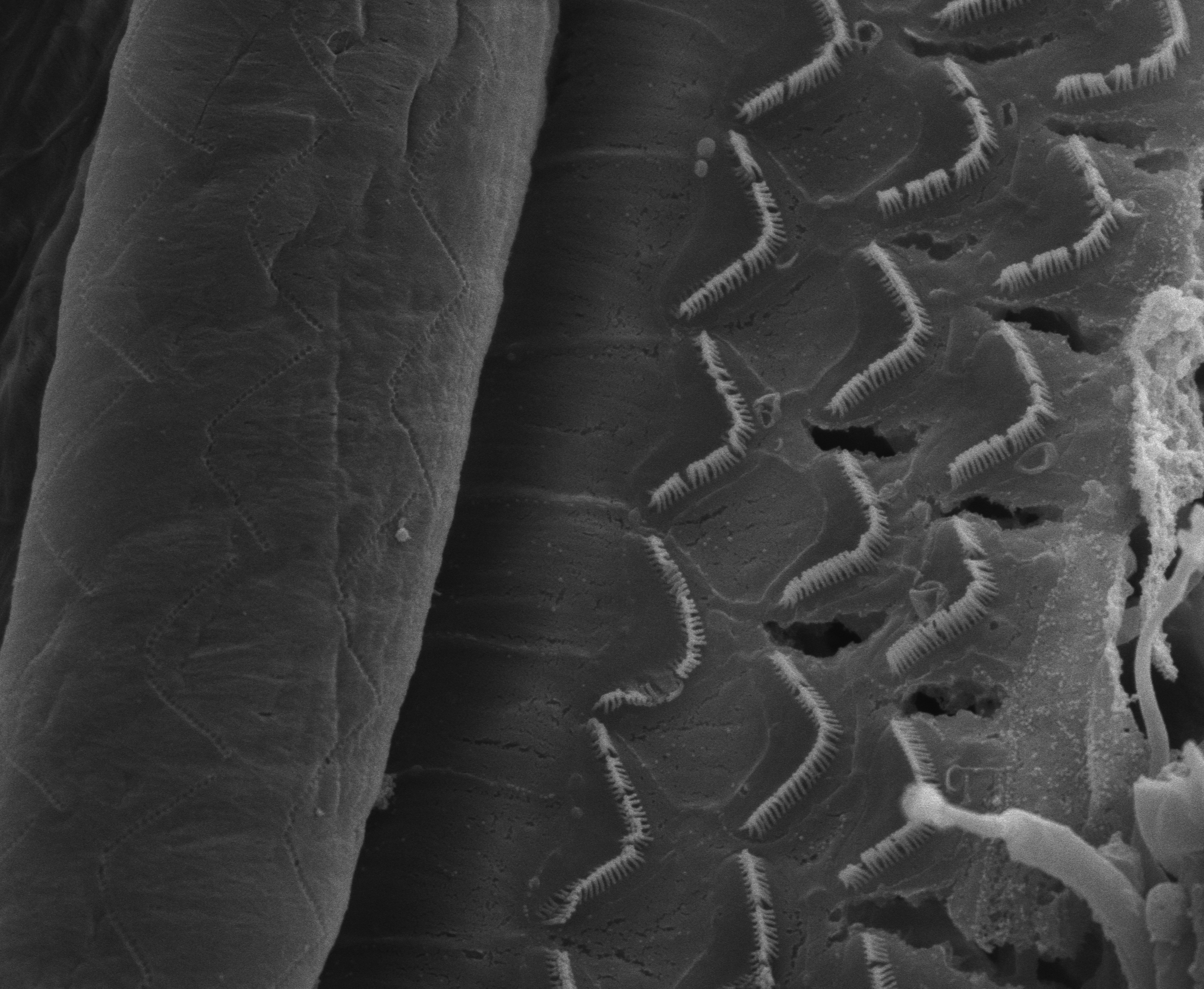
Outer hair cells of the mouse cochlea revealed by pulling back the tectorial membrane. Note the indentations on the membrane where the tips of the stereocilla were embedded.
Part of the mid-turn of the chinchilla cochlea revealing the orderly rows of inner and outer hair cells.
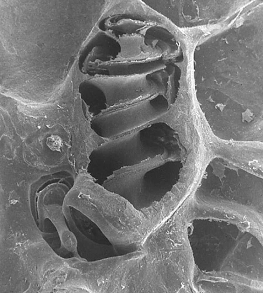
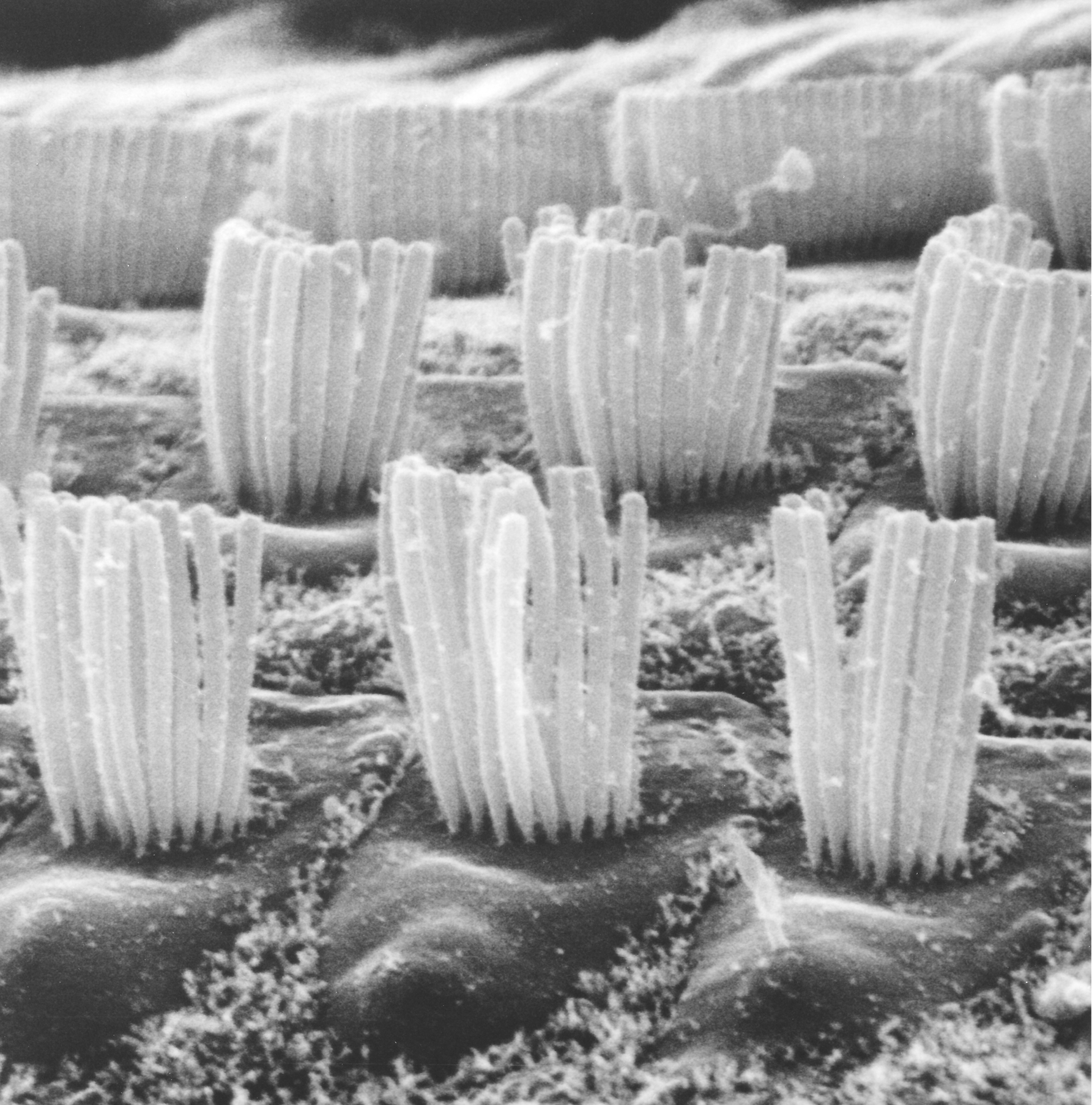
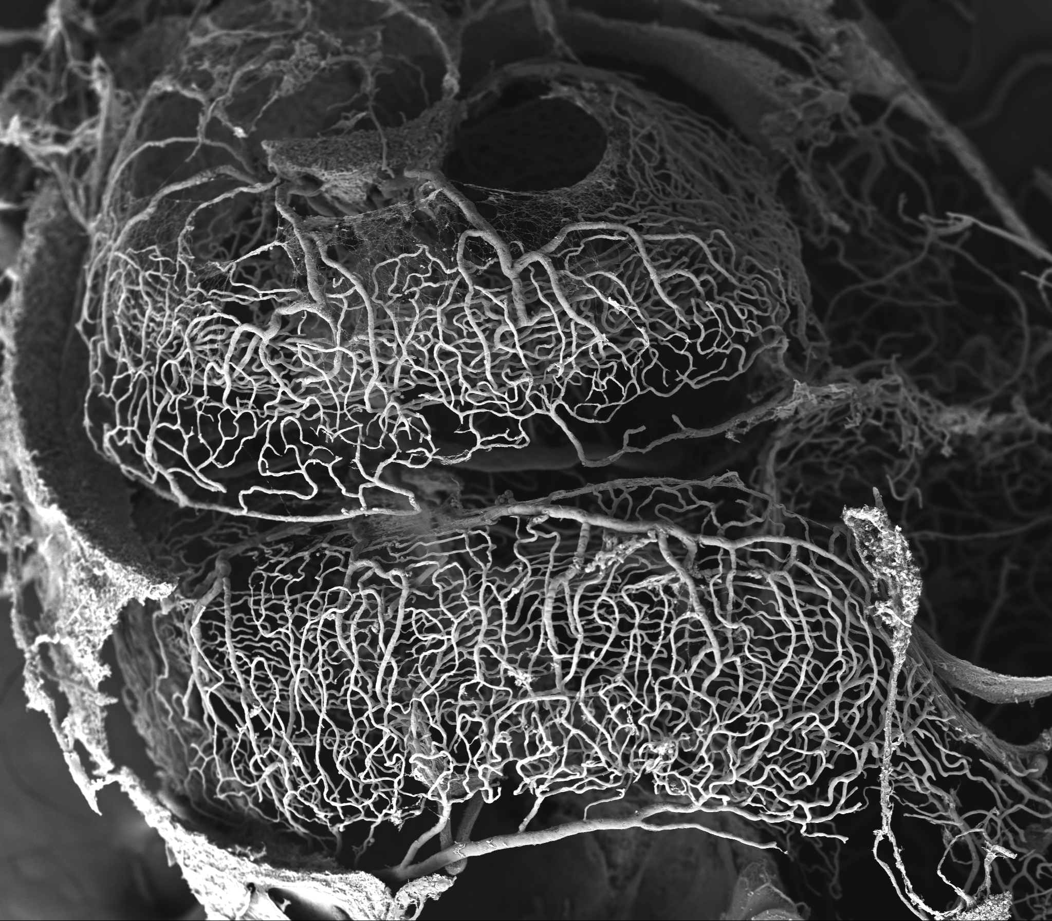
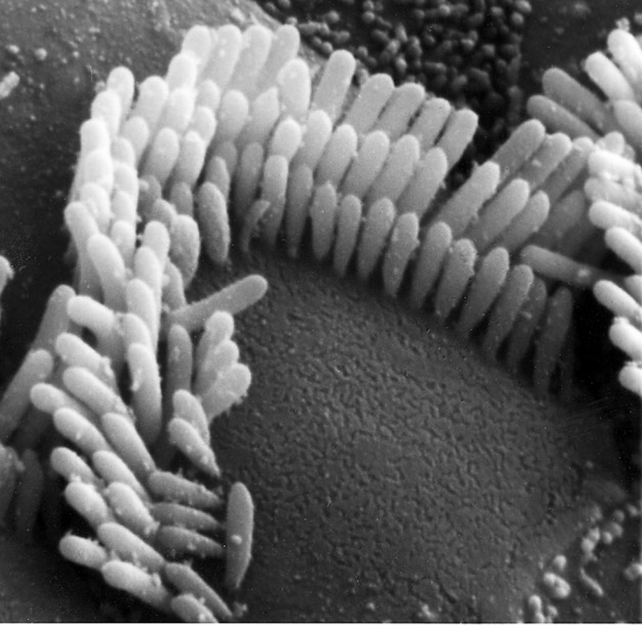
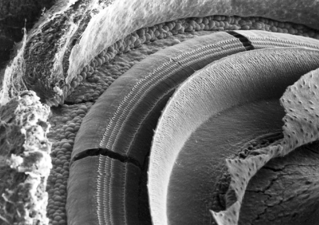
Image of the chinchilla cochlea after removal of part of its bony wall. Note the general spiral shape and at the cochlear base, the two “windows”: the oval window (containing the stapes footplate) and the round window.
High magnification image of outer hair cells at the apex of the (chinchilla) cochlea.
SEM of corrosion cast of the mouse cochlear vasculature. This image shows the capillary networks of the spiral ligament and the stria vascularis.
High magnification image of an outer hair cell after mild damage caused by acoustic trauma. Note the disarray of the stereocilla.
Transmission electron microscopy
Transmission electron microscopy (TEM) is a technique that captures an image of a sample by transmitting a beam of electrons through an ultra thin specimen sample. The electrons interact with the sample as they pass through to form the image. This allows capture of very high magnification images with high resolution of detail. Below are some of the images we have obtained using TEM in various parts of the inner ear. Click on any photo to obtain a higher resolution image that can be down loaded (copied or saved).
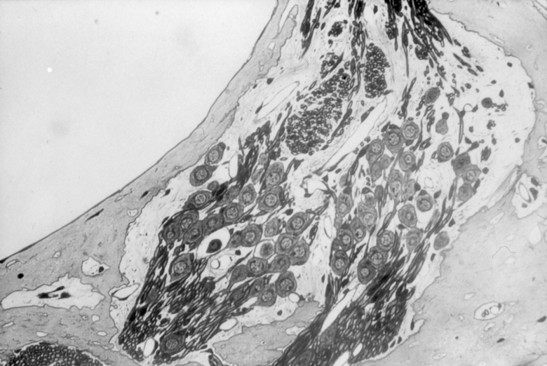
TEM image showing spiral ganglion cells that provide afferent and efferent connections to the hair cells in the organ of Corti.
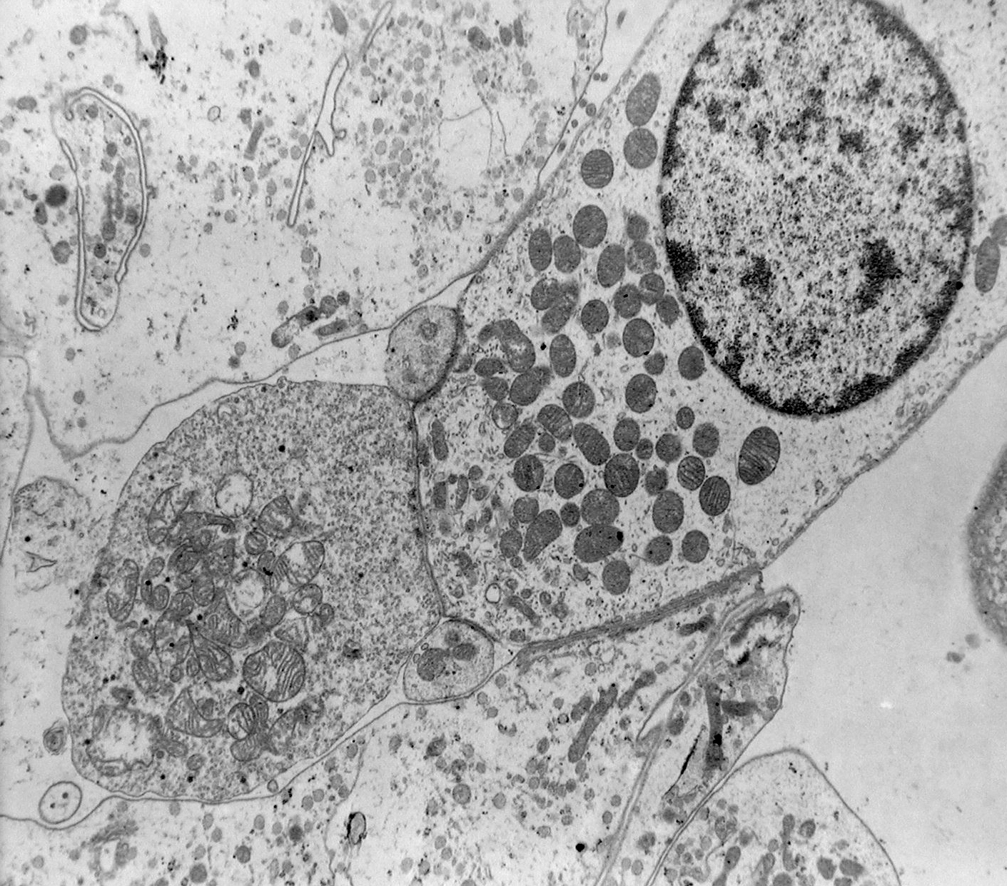
Image of the basal region of an outer hair cell with its nucleus and large number of mitochondria. In this basal area note both afferent and efferent synapses.
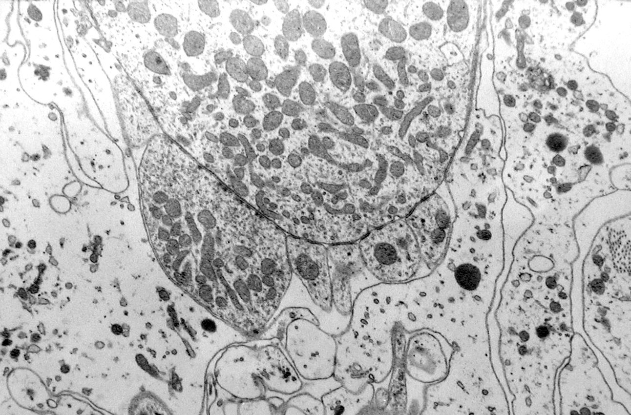
The synaptic region of an outer hair cell with its numerous mitochondria and nearby cochlear afferent and efferent synaptic terminals.
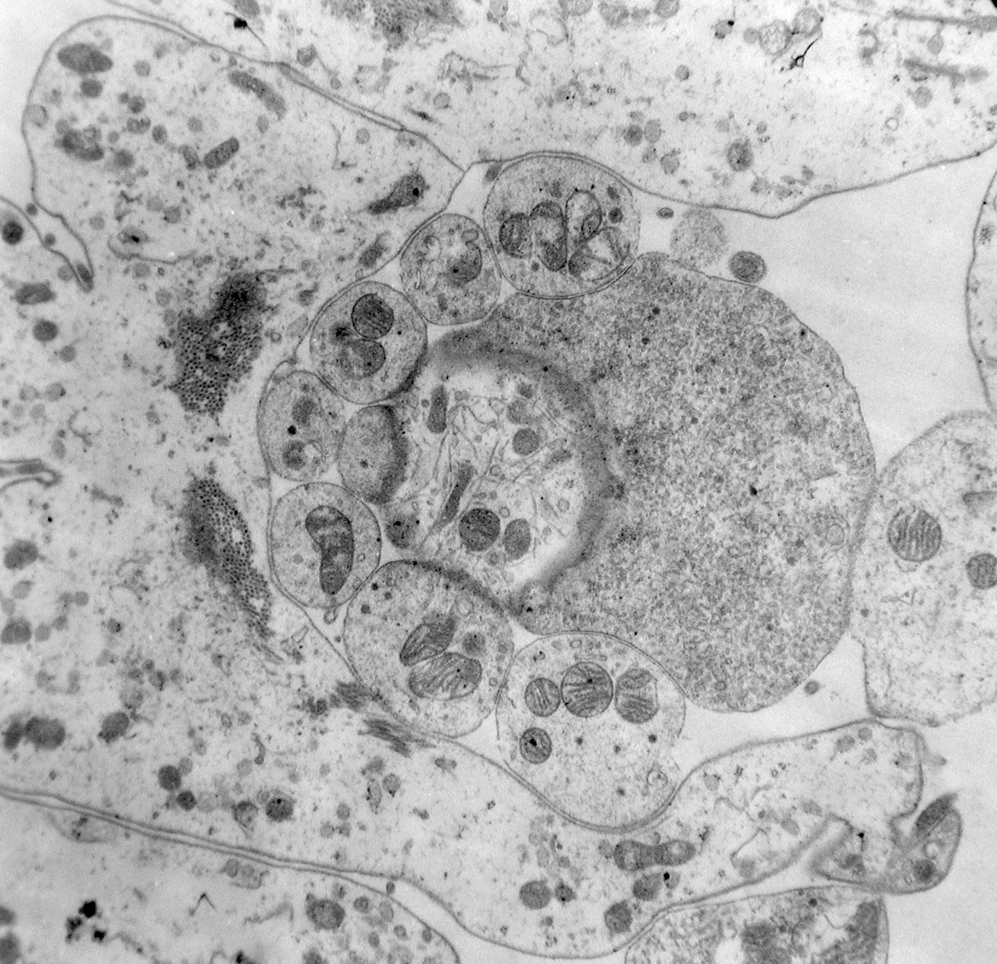
Cross-section through the basal area of an outer hair cell showing afferent and efferent synaptic connections.