The following pictures illustrate varying types of damage to the sensory cells of the cochlea caused by noise or drug exposure which result in a loss of hearing.
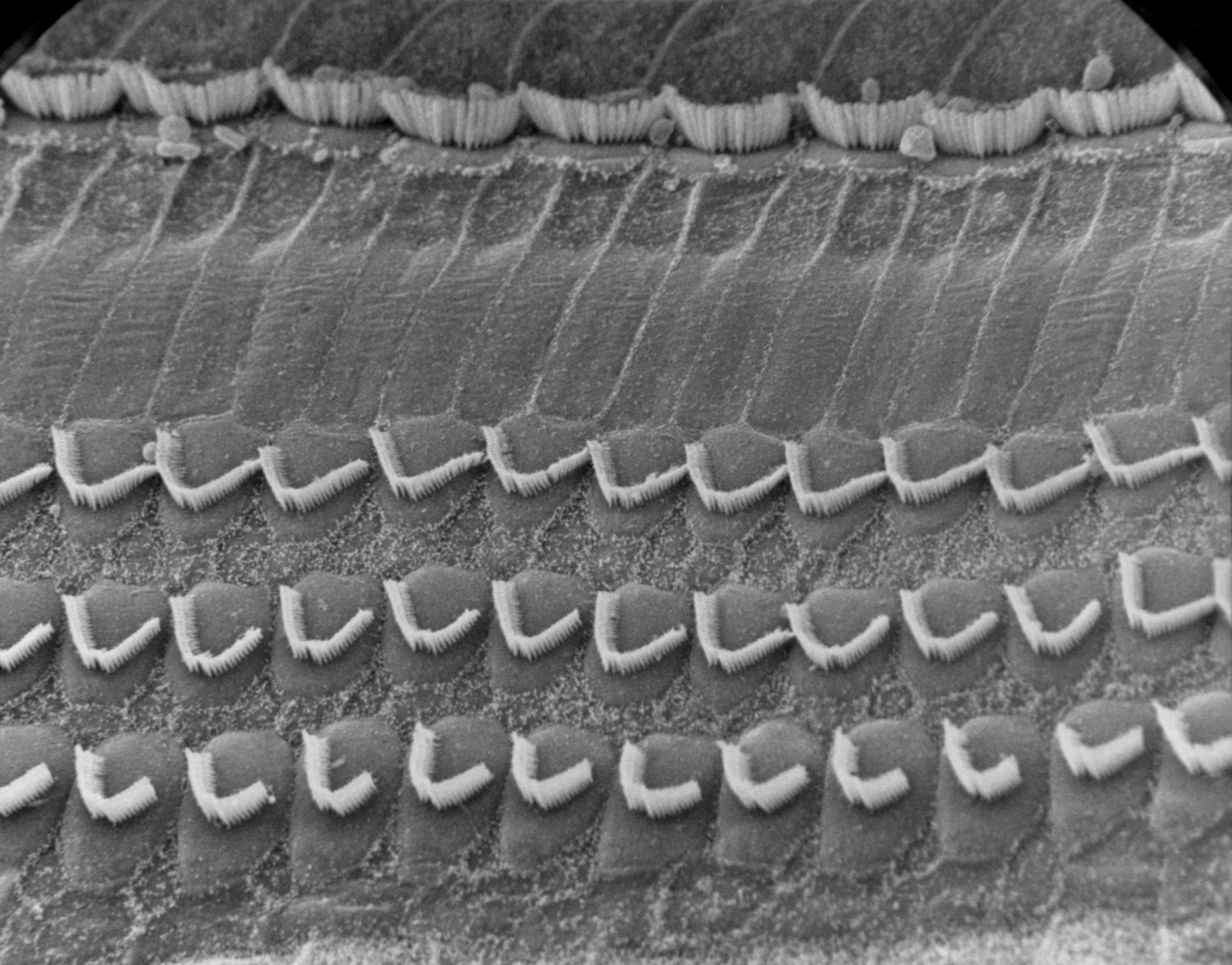
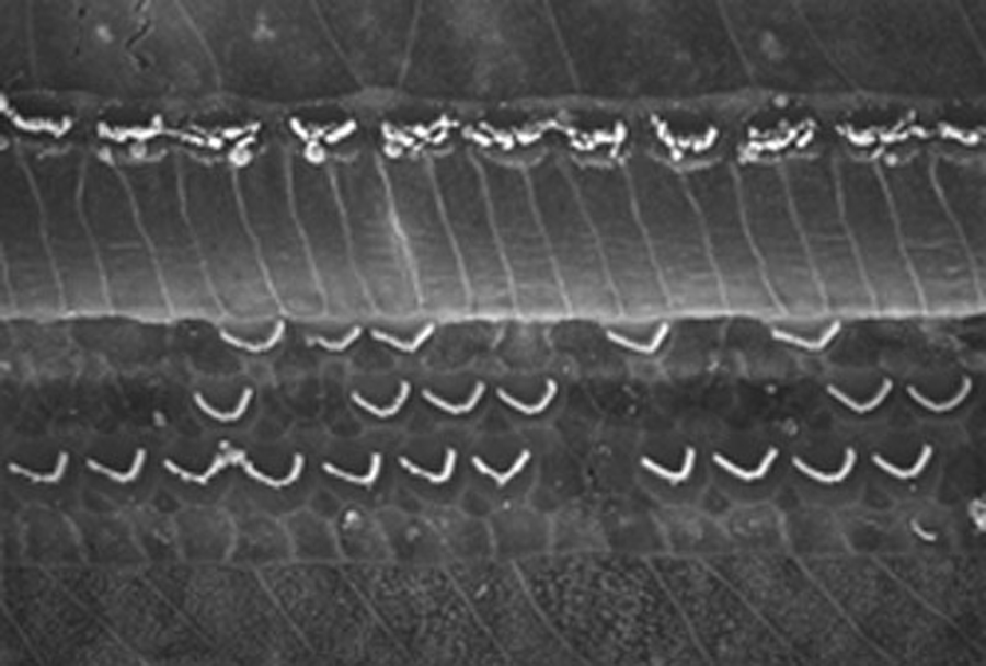
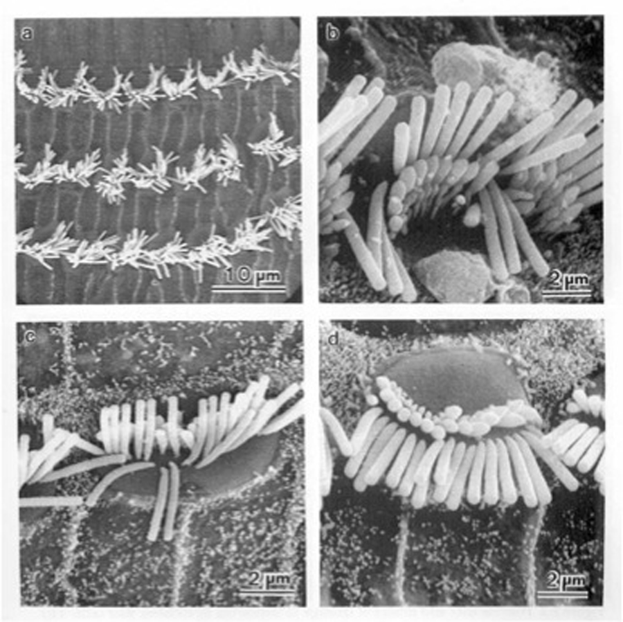
Sensory surface of the normal organ of Corti. Three rows of outer haircells and one row of inner hair cells are seen.
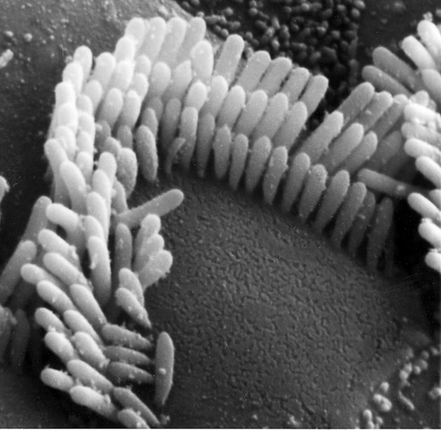
Mildly damaged outer haircell. Stereocillia are somewhat disarrayed, linkages between them are at least partially damaged impairing the function of the cell.
A mildly damaged inner haircell. Stereocillia are slightly disordered and several have fused together to form a “giant” stereocilium.
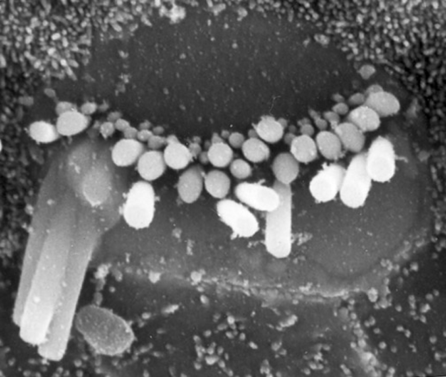
Severely damaged outer haircells. To the left a missing haircell has been replaced by ingrowing supporting cells. To the right are two haircells with stereocillia which are totally fused and/or disintegrating. It is highly improbable that these cells serve any active function.
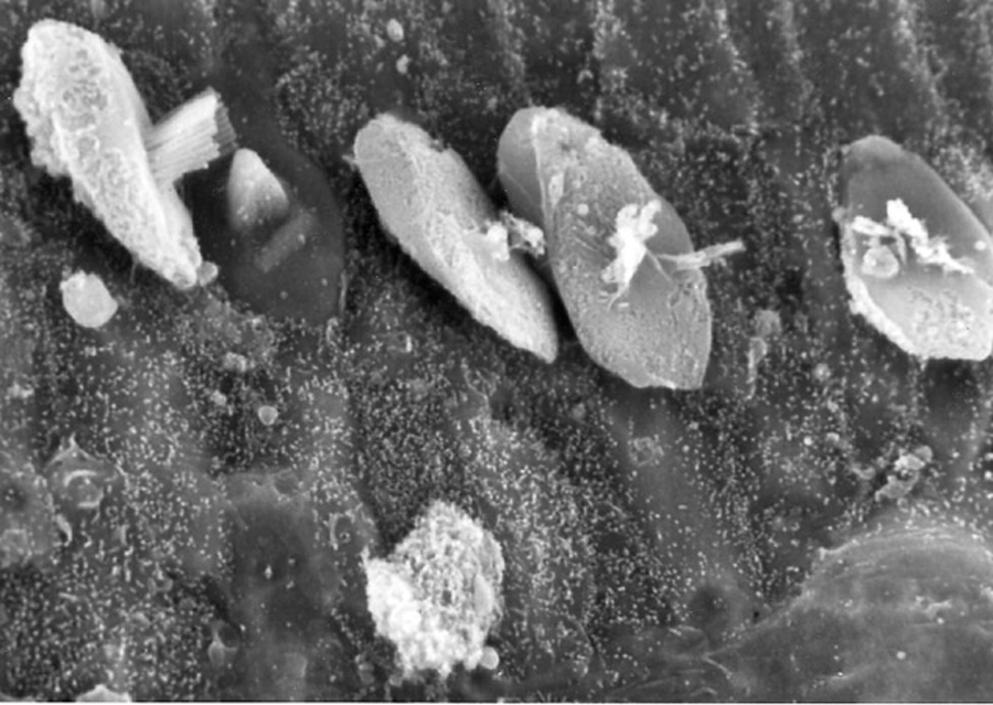
In these severely damaged outer haircells the cuticular plate, into which the stereocillia are anchored, has been ejected from the cell.
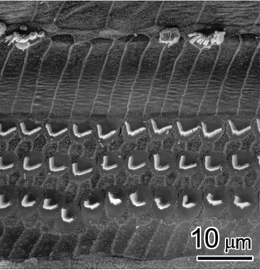
A severely damaged organ of Corti demonstrating an often seen sequence of haircell damage resulting from either noise trauma or ototoxic drugs.
First row outer haircells are most susceptible to many types of cochlea insult with second and third row outer haircells showing decreasing degrees of sensitivity.
Inner haircells frequently survive in regions of the cochlea devoid of any outer haircells. While this pattern of damage is common other patterns or a lack of pattern can often be found.
In this example, the organ of Corti was exposed to Carboplatin, causing the destruction of inner hair cells but leaving outer hair cells whole.
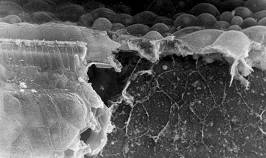
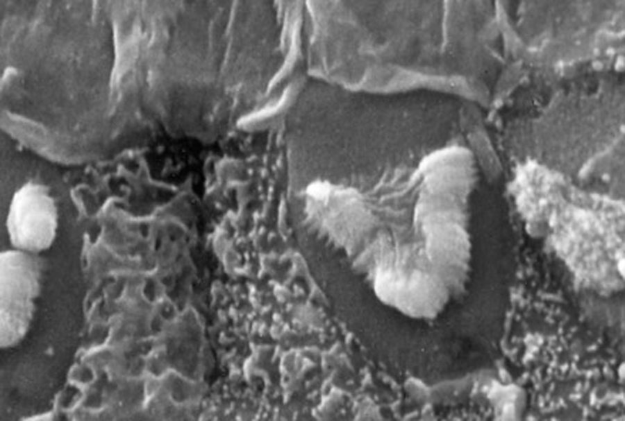
The edge of a lesion to the organ of Corti produced by severe sound trauma. To the left, some structure can be identified. Inner hair cells remain, however outer hair cells have been totally destroyed and their positions occupied by ingrowing supporting cells.
On the right of the micrograph only the basillar membrane can be seen, all organ of Corti structure has been destroyed.
This image shows varying degree of acoustic trauma on inner and outer hair cells. Stereocillia droop down and lose their rigidity, affecting the depolarization of hair cells and the efficient transition of sound waves to electrical activity in the brain.
References
Robert V. Harrison. The prevention of noise induced hearing loss in children – Review Article. Inter. J. Pediatrics. (2012) 2012: 473541.
Robert V. Harriosn. Noise-induced hearing loss in children: A ‘less than silent’ environmental danger. Paediatr. Child Health. (2008) 13(5): 377- 382.