We have utilized many different light microscopic techniques in our lab, including “regular” (broad spectrum) low and high power microscopes. With the use of fluorescent labeling our options have expanded. Here are some examples of cochlear anatomy investigated using confocal and laser sheet microscopes. All the images can be downloaded.
Laser thin sheet microscopy
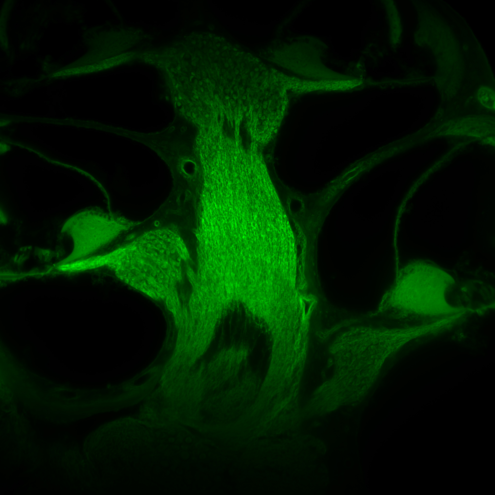
Cross-section through the cochlear modiolus (mouse model) using the laser thin sheet microscope. Spiral ganglion cell bodies, cochlear afferent axons and the organ of Corti can be clearly seen.
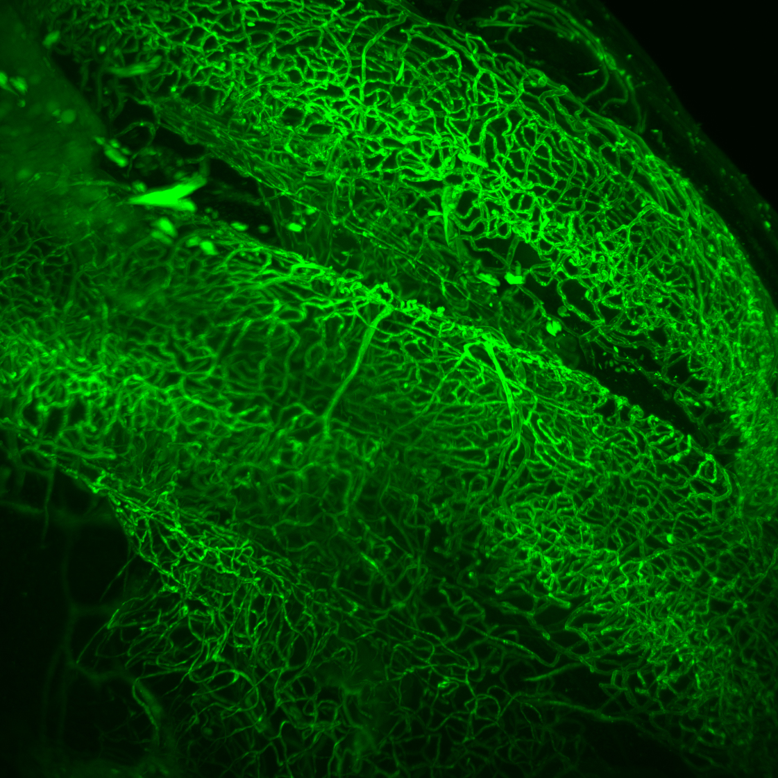
Image of the capillary network of the stria vascularis (mouse model). This image was made from a corrosion cast specimen using the laser thin sheet microscope.
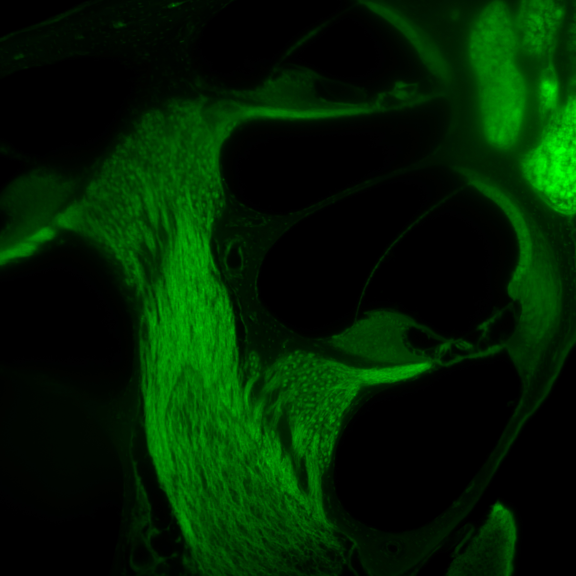
Lower turn of the mouse cochlea viewed with the laser thin sheet microscope. Note the organ of Corti and the stria vascularis on the border of scala media. Note also the clear resolution of cochlear neurons in the modiolus.
To see a video showing progressive cross-sectioning through the mouse cochlea, please click on ‘light microscopy’. The Z-stack was obtained using a laser thin sheet microscope.
To see a video showing a 3-D representation of cochlear vasculature (mouse model) please click on ‘partial cochlea rotation’. This is data obtained from a corrosion cast specimen using laser sheet microscopy.
Confocal microscopy
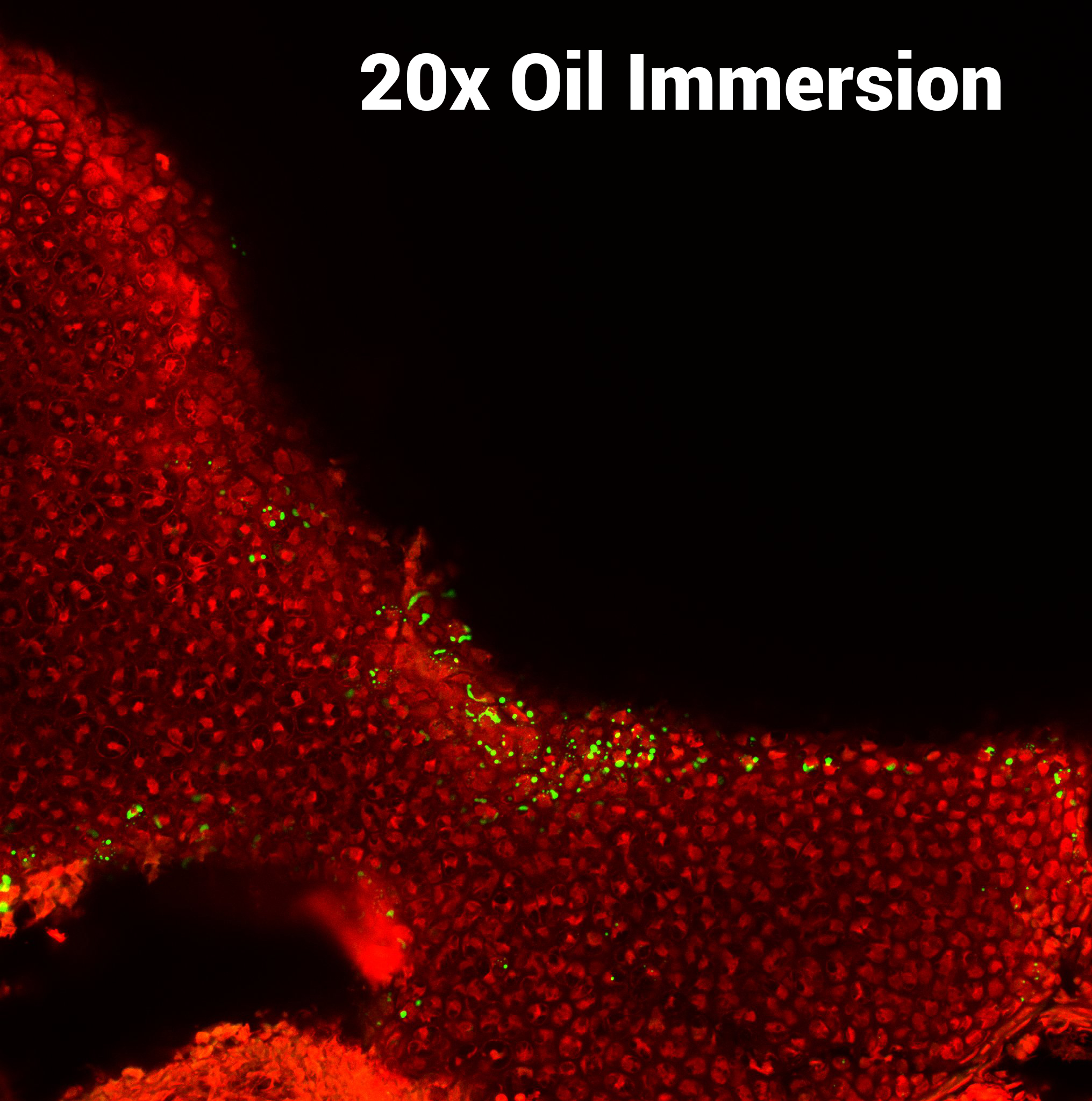
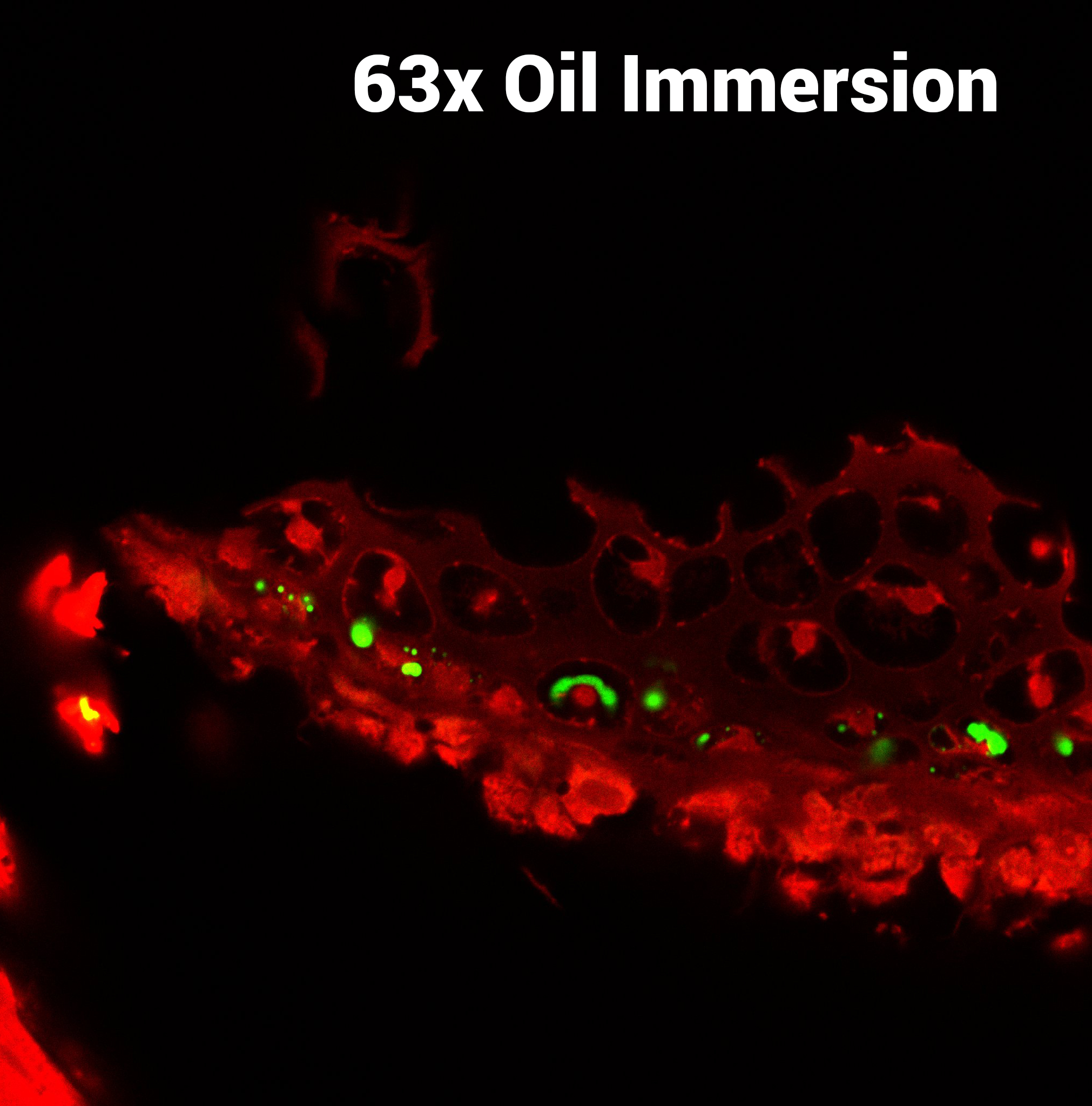
Image of a mouse cochlea infected with CMV virus. The viral particles are shown in green. This image was taken with a Nikon A1R Confocal at 20x resolution in oil immersion.
Image of CMV virus infection of the (mouse model) cochlea. The virus is shown in green. Image was taken with a Nikon A1R Confocal at 63x resolution in oil immersion.