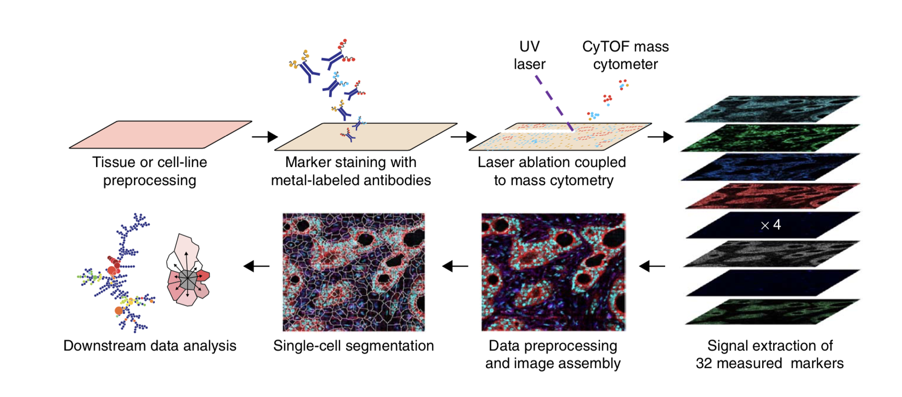

Imaging mass cytometry (IMC) enables deep, single-cell interrogation of cell phenotypes, cell-cell interactions and spatial proximity within normal and diseased tissues.
Hyperion can image tissue sections stained with up to 40 metal-containing reagents at 1 micron resolution, comparable to light microscopy, but with much higher content. Detecting markers with metal tagged Abs provides uniform staining with minimal background and no autofluorescence.
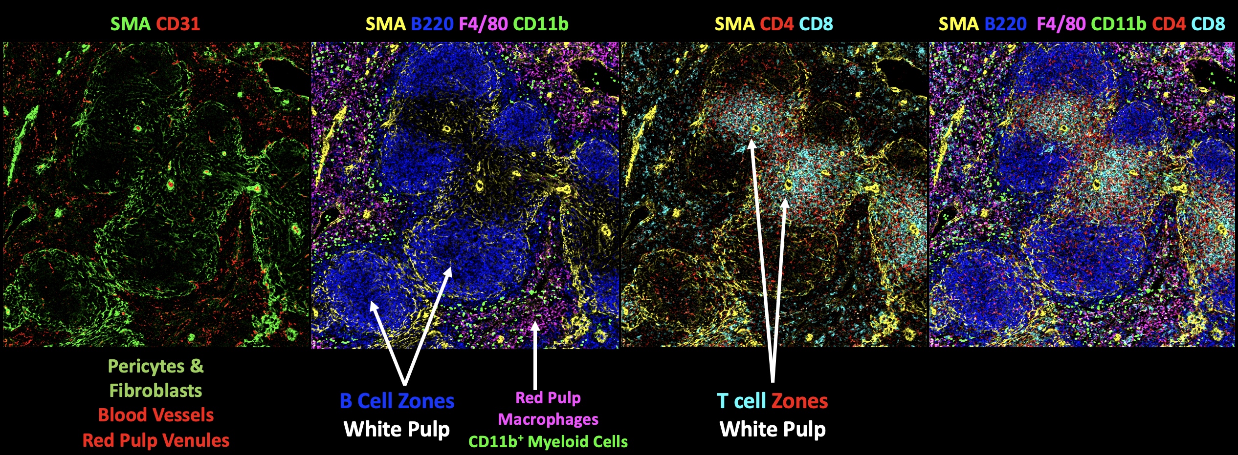
See examples of our IMC data for normal mouse tissues (spleen, thymus and small intestine), tumor immune profiling (mouse mammary gland and brain) and human tonsil.
We can perform small-scale IMC pilots on your sections cut from freshly frozen or formaldeyde-fixed, paraffin-embedded (FFPE) tissues. We will stain them with our validated metal-tagged IMC Ab panels for mouse frozen, mouse FFPE or human FFPE.
Contact Tina Chen with any questions, for a cost estimate and/or to schedule your pilot study.
IMC Services