We are located in the Echo Lab at The Hospital for Sick Children (SickKids), in section 4B of the Atrium.
Before the procedure, you will check in at the front desk. After getting your height and weight measured, you will be brought into our research room. Your research sonographer (the cardiac ultrasound technician who will be performing your test) will explain the procedure to ensure you understand the process and will answer any of your questions.
After obtaining your informed consent, you will be given a hospital gown to change into. The sonographer will ask you to lie flat on the hospital bed and attach three heart rhythm stickers to your chest, all while ensuring you are appropriately covered.
The sonographer will start the ultrasound by applying warm ultrasound gel on top of the ultrasound camera. Then, they will hold the camera to your skin on various areas of your chest: beside your sternum, on the side of your ribs, on your stomach and around your collarbone.This will allow the camera to view your heart from different angles. You may feel gentle pressure from the placement and movement of the camera. The sonographer will check in with you throughout the echo, but please speak up if there is anything you need.
Throughout the echo, you may be lying in different positions to help the sonographer obtain the best images your heart. You may need to lie on your left side, on your right side, flat on your back with a neutral head position and flat on your back with your head tilted up. Please inform the sonographer if any of these positions are challenging for you.
The echo test usually takes around 45-60 minutes and your parent or guardian will be allowed to stay with you throughout the procedure. The only thing you will need to do is to stay as still and relaxed as possible – this will help us get the best images of your heart! There is also a television to watch to help pass the time!
Watch Hermin ask Amanda, a sonographer, questions about echo:

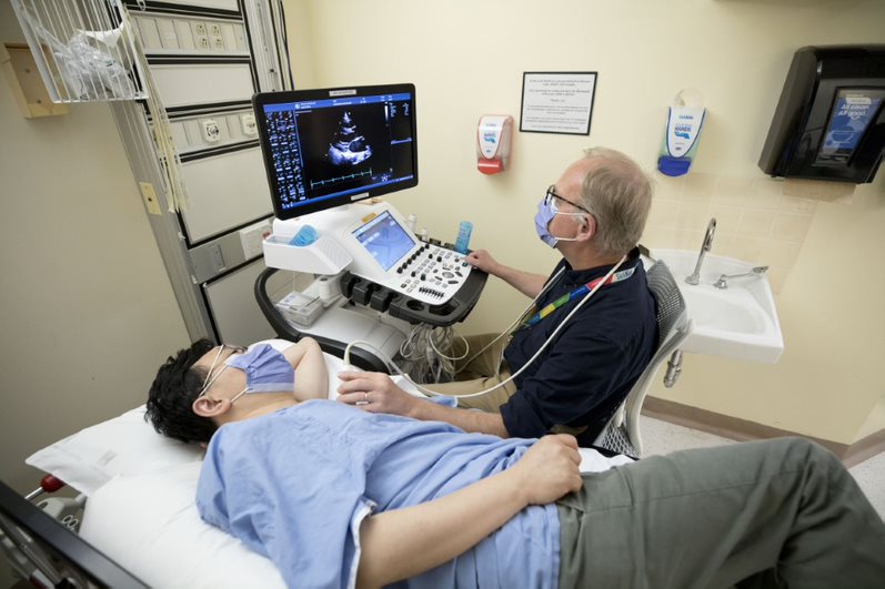
In this typical set-up for a cardiac ultrasound, the sonographer acquires images of the heart using the ultrasound machine, or “echo” machine in our dedicated research room.

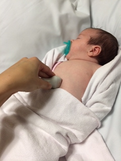
The ultrasound probe is positioned on the belly of an infant during a cardiac ultrasound, or “ECHO”.

