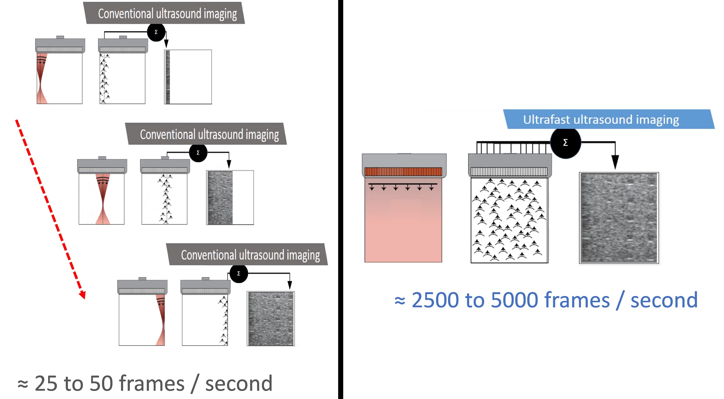
Ultrasound techniques currently used in echocardiography are limited by conventional frame rates. Ultrafast Ultrasound Imaging is able to capture images at frame rates up to 100 times faster than conventional imaging. Specific applications of this technology have been developed and tested for clinical use in paediatric and adult imaging.
Background
Although the use of ultrasonic plane-wave transmission rather than line-per-line focused beam transmission has been long researched, clinical application of this technology was only recently made possible through increasing computing capabilities and the introduction of powerful graphical processing unit (GPU)-based platforms, making it possible for the ultrasound system to acquire data in real-time.
Ultrafast Ultrasound Imaging is based on the use of plane or divergent waves, instead of focused beams. Plane and divergent waves are generated by applying a plane or circular delay on the elements of the transducer. In both cases, it is possible to reconstruct an entire image with a single emission. This imaging approach can result in lower spatial resolution and a lower contrast of the images. A technique that can be used to overcome this limitation is to use multiples plane waves transmitted at slightly different angles to obtain one image.
Coherent combinations of compounded plane-wave transmissions (also called coherent plane wave compounding) results in high-spatial resolution imaging, without compromising the high frame rate. As a result, blood and tissue movement and tissue structure can now be explored using high frame rate ultrasound imaging.

