PROJECTS


The Villemain lab runs a translational program. Starting with the development of a technology, the lab’s objective is to test its application and potential within a pre-clinical project (in vitro or animal model) and then to confirm its potential through clinical application studies. Click through the tabs below to learn more about how the Villemain Lab is using Ultrafast Ultrasound Imaging in different areas of medicine.

The team hopes to apply these new diagnostic tools in different areas of medicine to ultimately improve patient care.

Cardiology

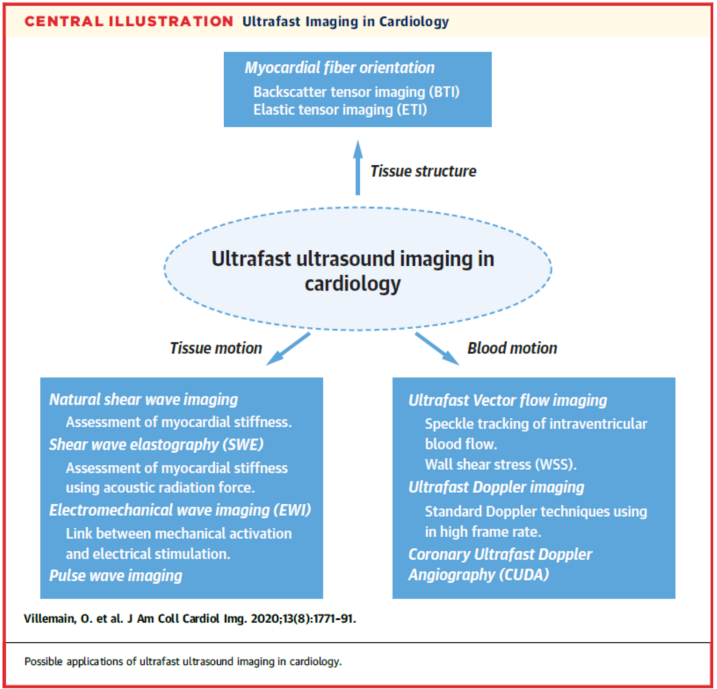
Specific applications of Ultrafast Imaging have been developed and tested for clinical use in paediatric and adult cardiac imaging. These include Ultrafast Doppler or vector flow imaging, shear wave imaging (assessment of tissue stiffness), electromechanical wave imaging and backscatter tensor imaging (assessment of fiber orientation).

Ultrafast ultrasound has great clinical potential in the assessment of cardiac function, in noninvasive hemodynamic analysis, while providing novel techniques for imaging coronary perfusion and evaluating rhythm disorders.

(figure from Villemain et al. JACC Cardiovascular Imaging, 2020)

Vascular

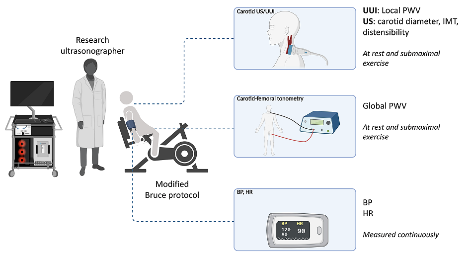
It is currently impossible to estimate local vascular stiffness despite the fact that this parameter is crucial for estimating the cardiovascular prognosis of certain patients. Ultrafast Ultrasound Imaging makes it possible to go beyond these limits.

Neurology

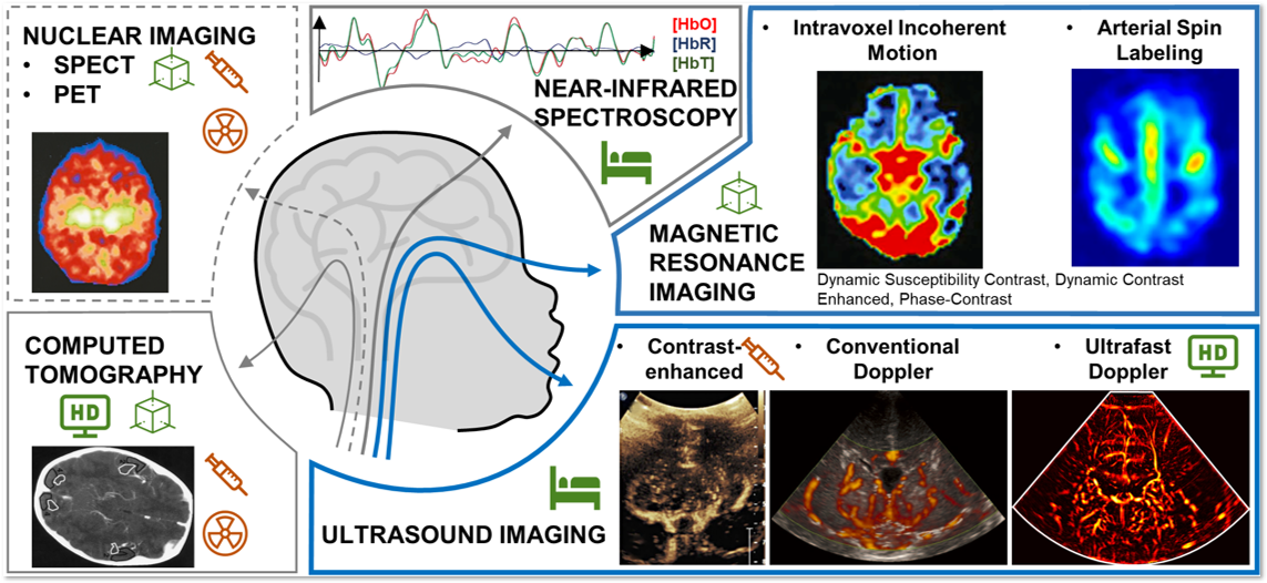
Ultrafast Ultrasound Imaging is able to produce images of the neonate brain vasculature with a high spatial resolution (down to 250 micrometers) and high temporal resolution (<1 millisecond). This allows the detection of many vascular abnormalities such as the beginning of an intraventricular hemorrhage. This imaging allows for the quantification of blood flow in the images, whereas conventional ultrasound scanners (the technology currently used in almost all clinical devices) can either display an image or quantify an area, but not both at once.

With Ultrafast Ultrasound Imaging, it becomes possible to follow fluctuations in blood velocity during the cardiac cycle, in both big arteries and small vessels. Many indicators can be derived from this information, such as the Resistivity Indexes ((Vmax – Vmin)/Vmax) which enables venous and arterial flow distinction and has been known to have strong diagnostic significance.

New emerging techniques also take advantage of the high spatial resolution produced using Ultrafast Ultrasound Imaging. This can help quantify a human’s vascular network in term of number of nodes and branches. Other features of Ultrafast Ultrasound Imaging such as Shear Wave Elastography can reveal tissue stiffness which can be another parameter of interest for diagnostic applications.

(figure from Baranger et al. NeuroImage Clinical, 2021)

Fetal and placenta explorations

Many pathological situations in the relationship between the fetus and the mother, such as intrauterine growth restriction or maternal hypertension, could benefit from a better understanding of placental perfusion and the difference between maternal and fetal blood flow. Ultrasfast Ultrasound Imaging brings new solutions and new perspectives for this.

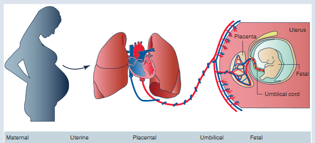
A diagram shows the lungs and heart of a pregnant woman, and how blood from the heart is pumped into the placenta. From there, the maternal blood flows into the fetus through the umbilical cord.
Placental perfusion, which is the perfusion of the placenta with maternal blood, is a fundamental aspect of the mother-fetus interaction