Skip to content
Taylor Lab
The Brain Tumour Group
Home
Medulloblastoma
Ependymoma
Team
Discoveries
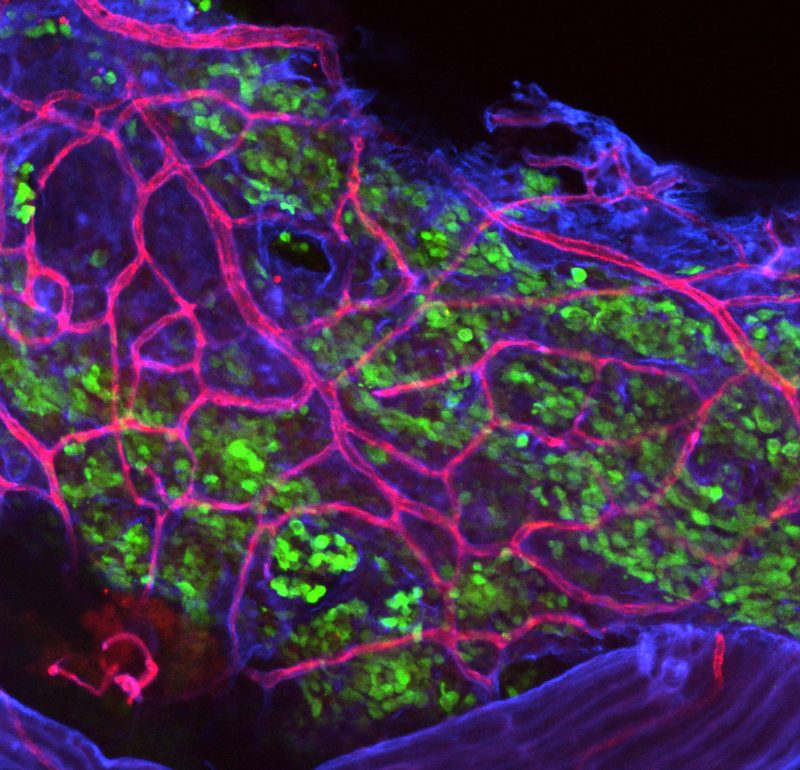
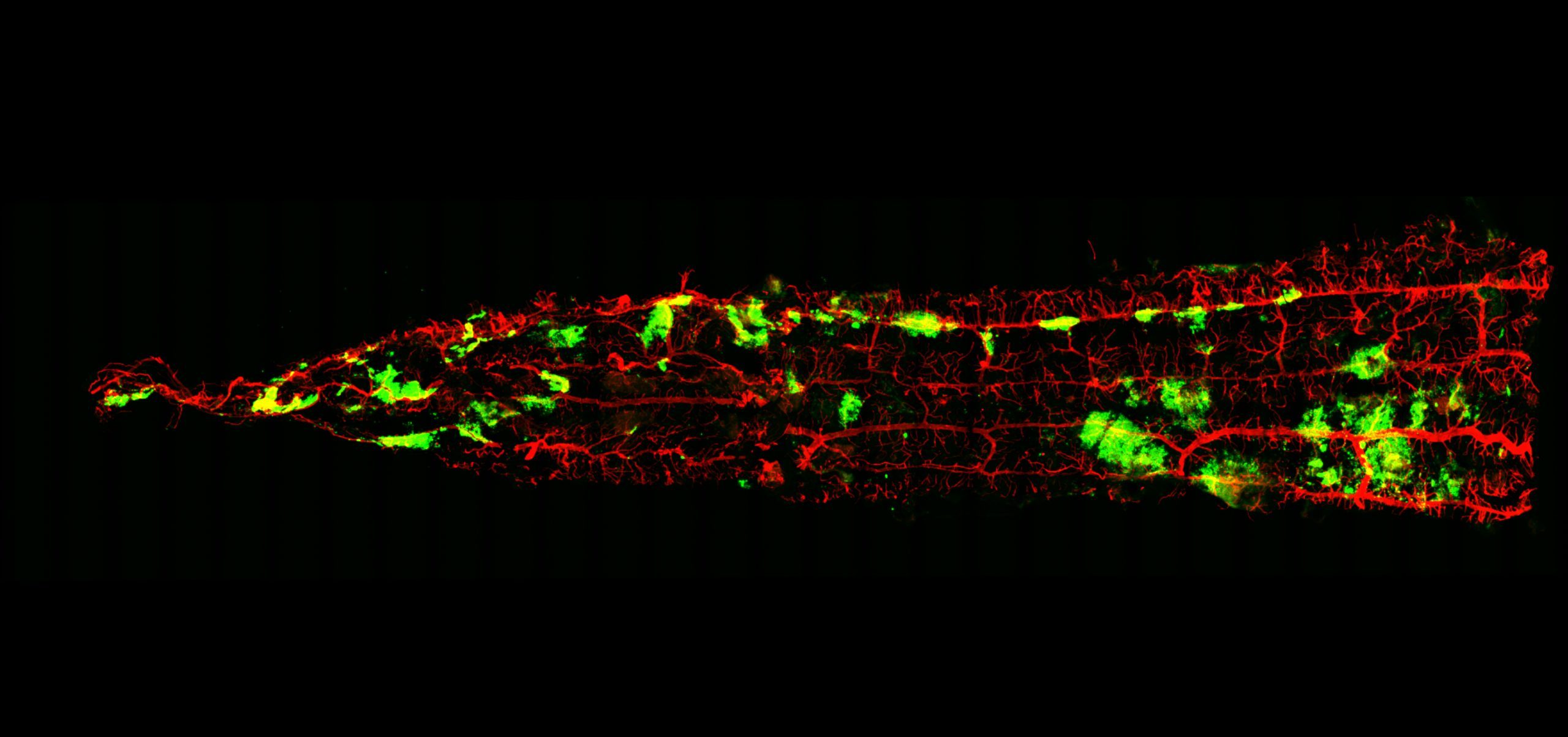
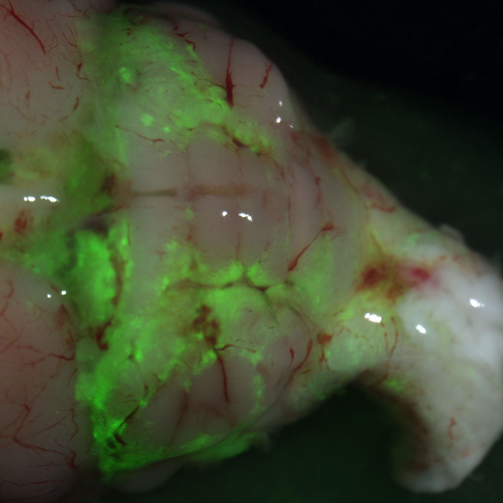
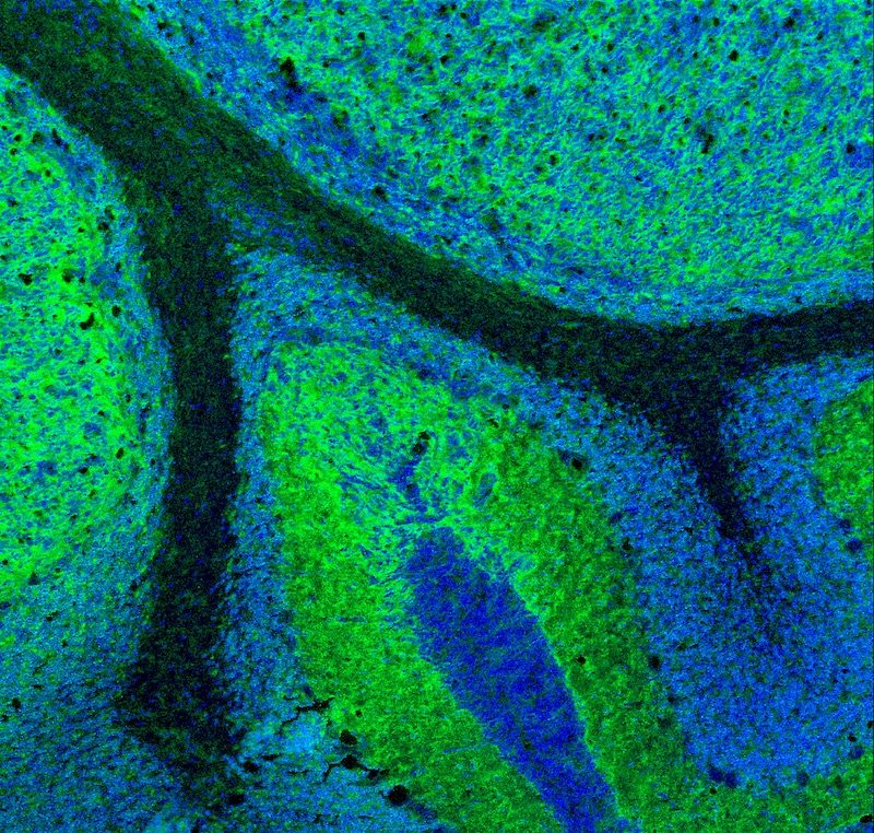
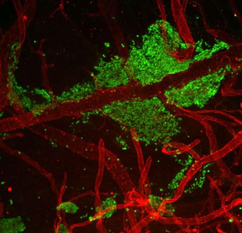
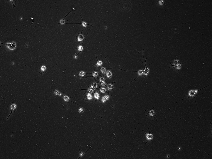
Gallery
Funding
Home
Medulloblastoma
Ependymoma
Team
Breakthroughs
Funding
Science
Fun
Science
Fun
Go to Top