Lung injury usually presents after trauma to the lung, for example radiation or prolonged mechanical ventilation. Our research focuses on the evaluation of structural and functional changes in the lung following lung injury.
Lung injury
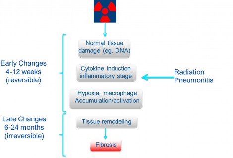
Diagram of the progression of radiation damage to the lung. Adapted from Ghafoori et al. Oncology Vol. 22 No. 1 (2008)
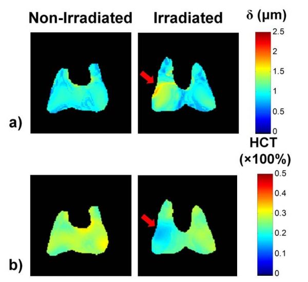
Radiation-induced lung injury
A common therapeutic approach for the treatment of cancers of the thoracic cavity is conformal radiation therapy (RT). Unfortunately, the lungs are a radiosensitive organ and are susceptible to damage from radiation, which may lead to Radiation-Induced Lung Injury (RILI) in some patients. RILI is a debilitating respiratory condition which can impact patient survival and post-treatment quality of life. Early detection of RILI is critical before the on-set of irreversible fibrosis. The Santyr lab is working with scientists and clinicians in the Department of Critical Care Medicine at SickKids, STTARR and the Princess Margaret Cancer Centre to develop and test MRI methods for early detection of RILI, particularly dissolved 129Xe imaging and apparent diffusion coefficient (ADC) mapping techniques.
Ventilator-induced lung injury
Ventilator-induced lung injury (VILI) is the most important factor limiting mechanical ventilation, the primary intervention for Acute Respiratory Distress Syndrome (ARDS). VILI can be caused by over-inflation of the lung, leading to an inflammatory response which, if unabated, progresses to fibrosis and ultimately organ failure. This is exacerbated by regional collapse of lung gas exchange units (e.g. atelectasis). One area of interest, in collaboration with the Kavanagh lab, is a novel method of reversing atelectasis called continuous negative abdominal pressure (CNAP). CNAP uses natural downward displacement of the diaphragm similar to normal breathing. 129Xe MRI can be used to detect atelectasis and subsequent recruitment using CNAP. The Santyr lab is working with scientists and clinicians in the Department of Critical Care Medicine at SickKids to develop and test MRI methods for monitoring the effectiveness of CNAP.
Bronchopulmonary dysplasia
Bronchopulmonary dysplasia (BPD) is the most common respiratory complication of pre-term birth. BPD can result when the premature infant is delivered before it can breathe on its own. Mechanical ventilation using oxygen, required for life support, can exacerbate BPD symptoms. BPD can lead to chronic respiratory diseases later in life, including asthma, chronic obstructive pulmonary disease and pulmonary hypertension. Hyperpolarized 129Xe MRI can potentially provide important regional information that can be used to detect BPD and guide treatment. The Santyr lab is working with scientists and clinicians in the Departments of Critical Care Medicine, Respiratory Medicine, Diagnostic Imaging and Neonatology at SickKids to develop and test MRI methods for diagnosing and monitoring BPD in newborns.