Hyperpolarized Gas MRI
MRI has been shown to be useful for providing excellent multislice soft tissue images in any arbitrary view. MRI use magnetism rather than x-rays, which means there is no radiation dose, especially important for children. Since conventional lung MRI is limited by poor signal, the Santyr lab is developing a new approach using an inhaled safe gas, hyperpolarized 129Xe, providing novel opportunities for imaging research. 129Xe is an inert contrast agent that can be visualized using MRI following inhalation, allowing structural and functional lung imaging.
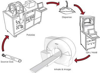
The Santyr lab utilizes research-dedicated MRI equipment within the Research MRI Facility at SickKids, to develop and implement the following 129Xe MRI lung imaging approaches in both children and animal models of disease.
Static ventilation imaging
Upon inhalation, 129Xe gas travels through the airways and fills the lung air spaces at which point an MRI image reflects the location of gas at that single point in time. Such “static” ventilation imaging shows the regional distribution of gas within the lung; areas within the lung that 129Xe cannot enter represent non-ventilated or regions of ventilation “defect”. The ventilation defect percent (VDP) is a useful measure for detecting and quantifying regional differences in lung function in diseases such as asthma and cystic fibrosis.
Apparent diffusion coefficient
The 129Xe gas present in the lung air spaces is free to diffuse around the airways and alveoli. Using diffusion weighted imaging one can sensitize the MRI signal to this diffusion to produce apparent diffusion coefficient (ADC) maps. Based on biophysical models of thermal diffusion of 129Xe atoms and measured ADC values one can then determine the structure of the airways of the lung and thus measure regional microanatomical alveolar size and shape of the airways. 129Xe ADC MRI can detect regional microstructural changes, including inflammation and emphysema due to injury.
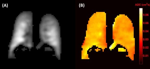
Single slice projection ventilation image (A) and corresponding ADC map (B) of a healthy paediatric volunteer.
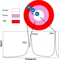
Top: Conceptual diagram of a single alveolus. The solubility of Xe in biological tissue allows for a small amount to exchange between the airspace, pulmonary tissue, and RBC compartments. Bottom: Xe in each of these compartments experiences a difference chemical shift in frequency due to the local environment, yielding the spectrum shown. Each signal peak is associated with a specific compartment shown in the diagram allowing for the probing of gas exchange as xenon exchanges between the airways and the blood pool.
Dissolved phase Xenon-129 imaging
129Xe gas not only fills the airways, but also dissolves into the surrounding lung tissue as well as the red blood cells (RBC) of the lung capillary bed. Within each of these three “compartments,” 129Xe demonstrates a different characteristic chemical shift which can be used to differentiate the MR signals emanating from each of the respective compartments. This makes 129Xe sensitive to changes in lung tissue and RBC compartment sizes as well as gas exchange between the air space, the lung tissue, and RBC pool. Gas exchange changes occur due to thickening of the tissue/gas barrier accompanying inflammation and fibrosis, hallmark features of many pulmonary diseases, such as lung injury.
Dissolved 129Xe also travels to organs beyond the lung, including the brain. In the brain, the chemical shift is different for the white matter, grey matter, red blood cells, and blood plasma. This allows 129Xe brain imaging to differentiate between these components in the brain. Furthermore, the chemical shift of the red blood cells depends on amount of oxygen in the blood. If the chemical shift can be quantifiably linked to the amount of oxygen in the blood, then it will be possible map the oxygen consumption in the brain. This can provide insight into many important physiological processes and diseases.
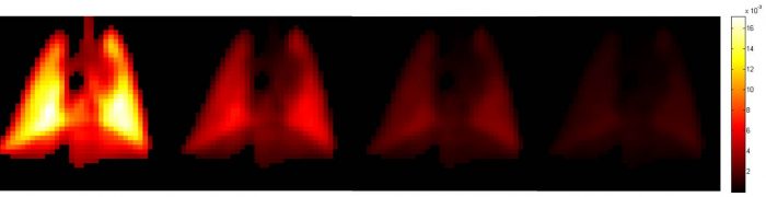
Multiple breath washout
Ventilation defects may not be static and as such, dynamic lung imaging methods must be developed. After inhaling a single dose of hyperpolarized 129Xe, MRI can be performed during successive breaths of room air in order to monitor the washout of the 129Xe gas. The resulting images can be used to calculate the fractional ventilation parameter, which provides information that is similar to VDP, but independent of the absolute 129Xe signal which can be affected by MRI system-dependent parameters.