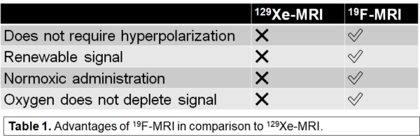
Fluorine-19 is a versatile, nuclear magnetic resonance active nucleus that has potential in imaging-based cell tracking, lung imaging, and drug development. Fluorinated contrast agents such as perfluoropolyether (PFPE), perfluoro-15-crown-5-ether (PFCE), sulfur hexafluoride (SF6), perfluoropropane (C3F8), and hexafluoroethane (C2F6) can be used to overcome limitations presented in hyperpolarized gas MRI.
Fluorine (19F) MRI
Inert Fluorinated Gas MRI
Similar to hyperpolarized 129Xe MRI, an emergent technology for lung imaging involves the use of inert fluorinated gases to measure lung ventilation. Fluorinated gases such as perfluorpropane (PFP; C3F8), are detectable with MRI, and are safe, non-toxic, and relatively inexpensive. Most importantly, fluorinated gases do not require hyperpolarization and the associated costs or equipment, in order to be detectable with MRI. Therefore, inert fluorinated gas MRI has significant potential as a lower cost alternative to hyperpolarized MRI for lung imaging. In addition, these gases can be freely administered in normoxic mixtures (21% oxygen), allowing for extended, free-breathing characterization of gas kinetics (such as gas wash-in or wash-out) in a manner that is not possible with hyperpolarized gases.

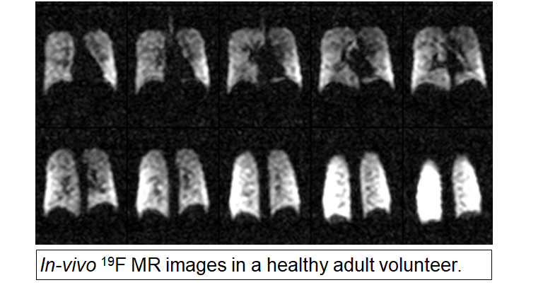
Our group has recently demonstrated 19F-MRI in a healthy adult volunteer with perfluoropropane (PFP) gas (79% PFP, 21% O2). This technology may also be used in an MBW fashion to improve investigation of gas kinetics.

Fluorine-19 MRI in cell tracking/imaging
Cell tracking is an important tool for the field of cellular therapy. The goals of cellular therapy are to stimulate the immune system to elicit an immune response and regenerate damaged tissues using stem cells. The requirement and need to track cells noninvasively is of great importance for the development of cellular therapies for eventual clinical translation and acceptance.
Fluorinated contrast agents and 19F MRI have emerged as a potential technique for noninvasive cell tracking experiments. Cells can become NMR sensitive by labeling with fluorinated compounds and delivered to the lungs. Their biodistribution can then be imaged using 19F MRI and activity of the cells can be monitored. One of the advantages of using 19F MRI is that, unlike other negative contrast agents used for cell tracking, 19F MRI gives positive signal images. In addition, there is no background signal coming from the inherent lack of fluorine signal in the lungs. 19F MRI can allow for direct quantification of fluorine-labeled cells, which makes it a powerful imaging modality in cell tracking. 19F MRI, however, does not give any structural information of the lungs, so proton MRI is often obtained in parallel to overlay with the 19F images.
