Cystic fibrosis (CF) is the most common genetic disease affecting children and young adults in Canada, caused by defects in the CFTR encoding gene. CF causes lung obstruction making it difficult for the child to breath. Currently used diagnostic tools for CF are insensitive to regional disease and are highly variable, especially in children. The Santyr lab is working with scientists and clinicians in the SickKids CF Centre to develop and test MRI methods for measurement of regional disease in children afflicted by CF.
Cystic fibrosis
Lung clearance index
The lung clearance index (LCI) is a novel method for measurement of lung physiology, derived from inert gas multiple breath washout (MBW) test. It is performed during quiet breathing by the child and thus is less variable than traditional pulmonary function tests involving forced expiration maneuvers. LCI has also been shown to be feasible in infants. LCI reflects global ventilation inhomogeneity and is a highly sensitive marker of early obstructive lung disease. This project is investigating the correlation between LCI and quantitative regional analysis of 129Xe MRI in pediatric healthy volunteers and patients with CF.
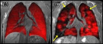
HP 129Xe MR images overlaid on conventional MR images obtained in our laboratory at SickKids from (A) a healthy volunteer and (B) a patient with cystic fibrosis. Arrows indicate areas of ventilation defect.
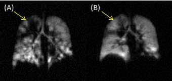
Hyperpolarized 129Xe MR images acquired in a paediatric patient with cystic fibrosis immediately following the diagnosis of a pulmonary exacerbation (A) and after 2 weeks of treatment with antibiotics (B). Arrows indicate area of improved ventilation post treatment.
Pulmonary exacerbation
Patients with CF experience periods of worsening symptoms known as pulmonary exacerbations that make day-to-day living a challenge and can result in irreversible lung damage. It is particularly important to treat an exacerbation quickly and effectively, especially in children. 129Xe MRI has been shown to be very sensitive to CF exacerbations. This project uses 129Xe MRI to monitor the response of CF exacerbations following treatment with conventional (i.e. antibiotic) as well as new medications.
Multiple breath washout
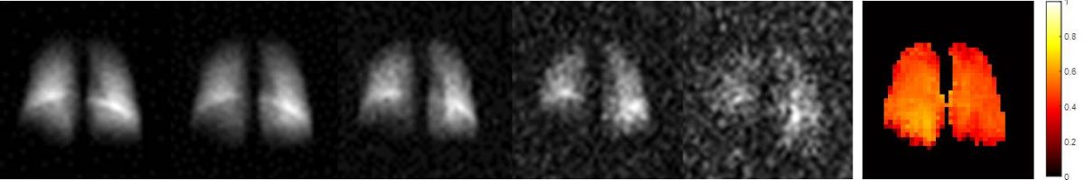
LCI is emerging as a sensitive indicator of ventilation inhomogeneity. Unfortunately, LCI is a whole lung measurement performed at the mouth, and it is unable to localize the disease within the lungs. Multiple breath washout hyperpolarized 129Xe imaging can be used to calculate regional ventilation maps, information that is similar to LCI, except with the added benefit of regional information. With this sensitive, regional information, we will be able to localize air trapping and quantify the poorly ventilated regions of the lung. One of our studies seeks to optimize regional ventilation mapping while also observing whether it correlates with LCI in patients with CF.