Genome organization is important for the regulation of genes and cell fate determinations. For gene expression to occur, regulatory elements come into physical proximity of genes. Rearranging these gene regulatory networks and genome organization alters developmental programs and causes rare diseases and cancer.

Various disease-associated protein-coding genes have been identified. However, coding genes represent only ~2% of the transcribed human genome, while > 95% consists of transcribed non-coding DNA. The majority of disease-relevant genetic variations and mechanisms reside in the non-coding genome, including long non-coding RNAs (lncRNAs) which do not encode for proteins. Intensive research demonstrates that lncRNAs and noncoding DNA are integral to the function of cells, particularly in the control of gene activity and in the maintenance of genome organization, a discovery revolutionizing our understanding of biology and of pathogenesis.

To better understand biology and etiology of disease, we need to determine how the non-coding genome functions and how it impacts normal and disease states. The Maass Lab studies molecular mechanisms associated with the non-coding genome.

Inter-chromosomal contacts

Decoding the functions of inter-chromosomal contacts

Intra-chromosomal organization within chromosomes is well understood; however, inter-chromosomal interactions between different non-homologous chromosomes also exist, but they are less studied and the molecular mechanisms remain elusive. Thus far, only a few studies highlight that inter-chromosomal contacts are crucial for development and gene regulation, and they are implicated in disease. The Maass Lab investigates the inter-chromosomal contact between a long non-coding RNA (lncRNA) locus and a developmental morphogenesis gene which is proof-of-concept that inter-chromosomal contacts drive gene regulation, and if replaced, such loci can be associated with disease.

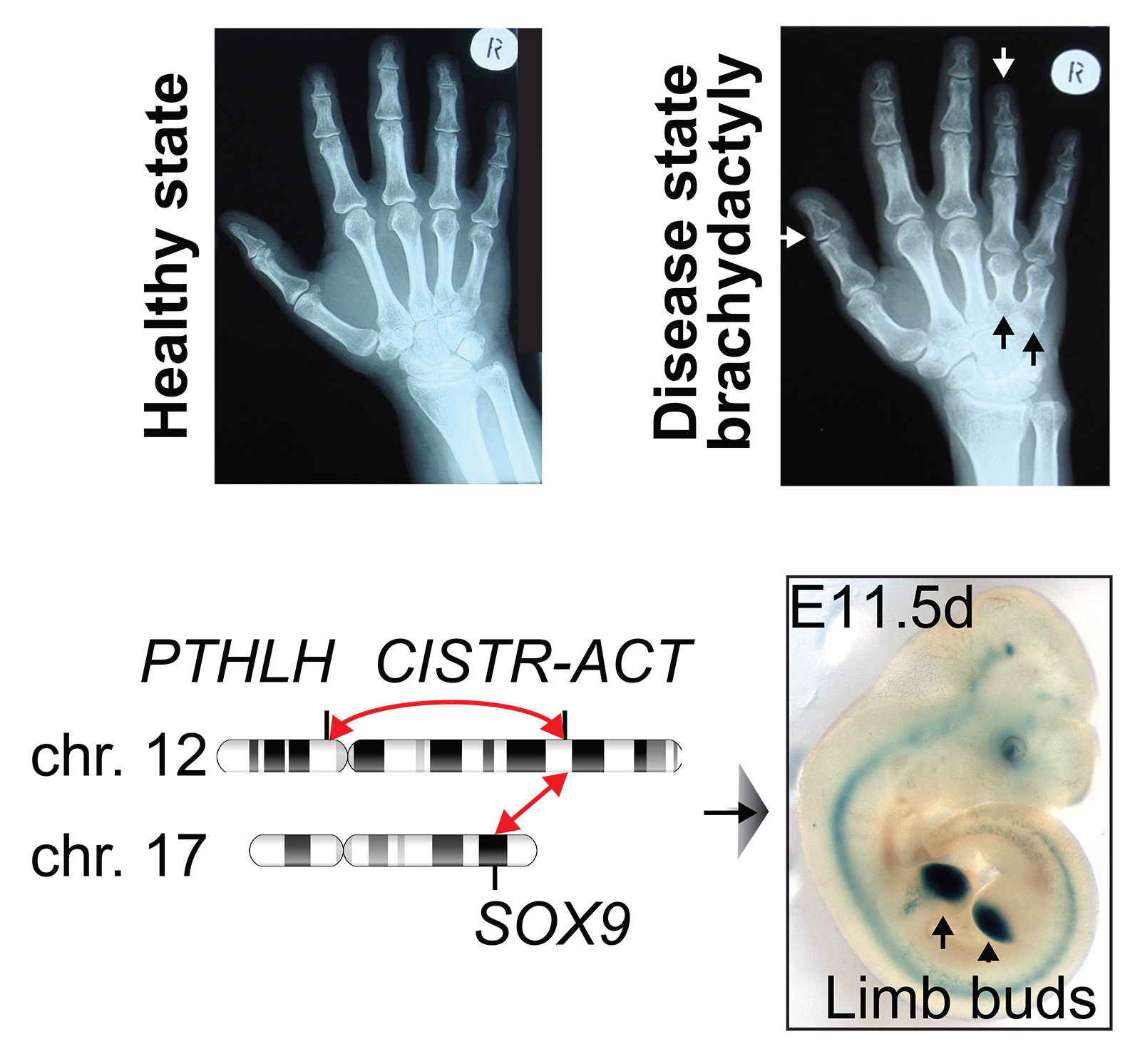
Specifically, the human lncRNA locus CISTR-ACT, located on chromosome 12 facilitates an inter-chromosomal contact with the transcription factor SOX9 on chromosome 17. Disrupting CISTR-ACT’s regulatory landscape caused the congenital cartilage malformation brachydactyly type E (shortened fingers and extremities) by misplacing the CISTR-ACT locus to a derivative chromosome.

Hand roentgenograms show shortened fingers (brachydactyly), caused by misplaced inter-chromosomal interactions between the long non-coding RNA gene locus CISTR-ACT (chromosome 12) and the transcription factor SOX9 (chromosome 17).

Live-Cell Imaging

Towards a better understanding how genomic loci interact in living cells

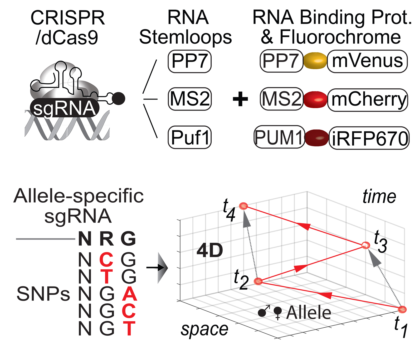
With the advent of the CRISPR/Cas9 system, any genomic locus can be targeted and labeled in live cells. Introducing CRISPR live-cell imaging (CLING) overcame the limitation of studying genomic interactions in fixed cells. CLING technology leverages inactive dCas9 together with multiplexable RNA-aptamer stem loops attached to the sgRNA that recruit aptamer-binding moieties fused to fluorescent proteins to target genomic loci.

Distinguishing between parental alleles to investigate allele-specific effects (maternal/paternal) has been a major challenge and has not been possible in living cells so far. To further improve live-cell imaging and to enable studying genomic interactions of the parental alleles in living cells, SNP-CLING exploits heterozygous single nucleotide polymorphisms (SNPs) in the protospacer adjacent motif (5’ – NRG – 3’) which is needed for dCas9 to target a genomic locus. The Maass Lab is equipped with the ultra-sensitive Airyscan FAST imaging system to explore the properties of genomic loci and alleles in living cells across space and across time.

CRISPR/dCas9 live-cell imaging (CLING) uses RNA binding proteins (MS2, PP7, Puf1) fused to fluorescent reporters to target DNA loci.

Genomics

High-throughput characterization of gene-regulatory elements

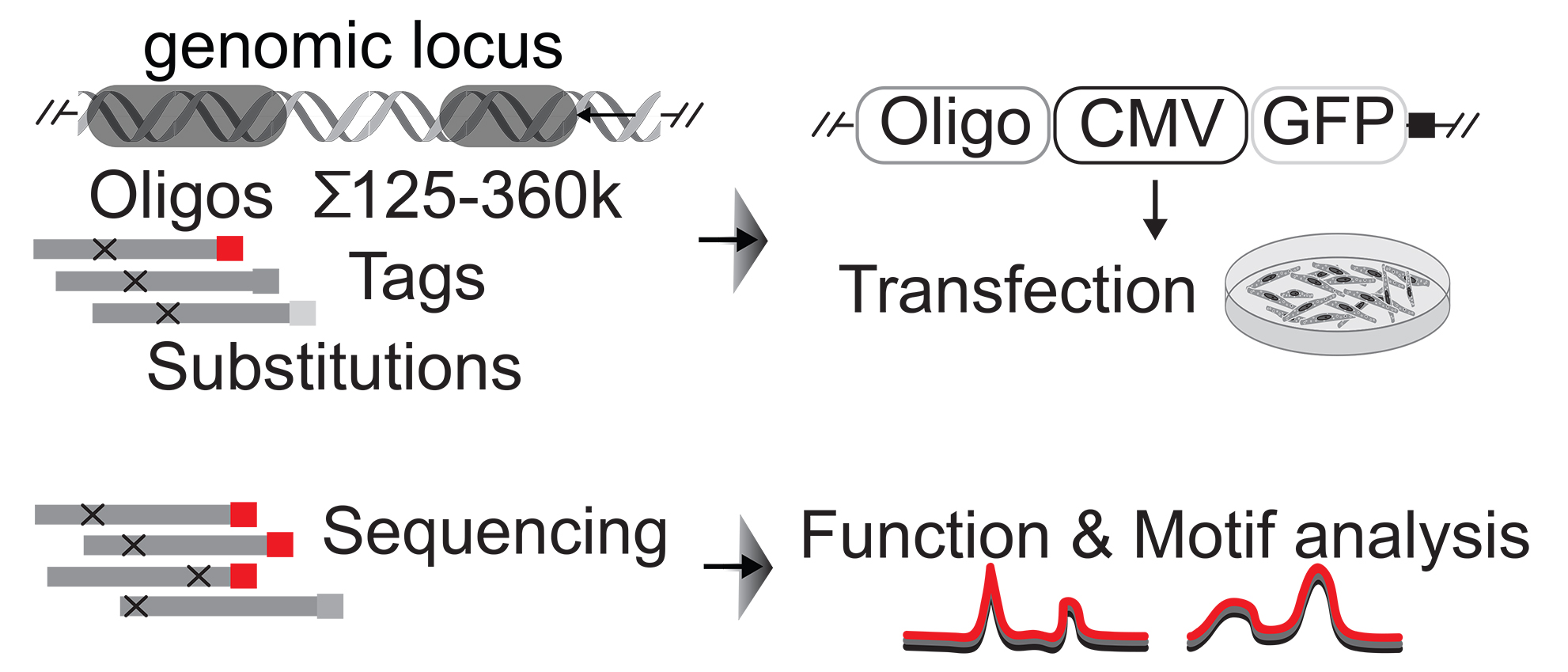
The functionality of a genomic region can be determined by reporter assays, where the reporter’s transcriptional activity is proportional to the tested candidate region. To assess transcriptional activity and regulatory motifs on global scales, massively parallel reporter assay (MPRA) technology has been introduced by testing thousands of genomic regions simultaneously. We apply this technology to identify regulatory features of genomic non-coding regions, such as which transcription factor binding motifs regulate tissue-specific expression of a lncRNA. Moreover, we decode the non-coding genome by RNA and ChIP-seq, chromosome conformation capturing, and MPRAs.

Massively parallel reporter assay (MPRAs) leverage thousands of oligonucleotides to determine regulatory DNA sequence elements in a genomics high-throughput approach.