Working at both ends of the cell size scale, our work relies heavily on state-of-the-art imaging techniques.
Several images from our lab have featured on journal and book covers, and won awards:
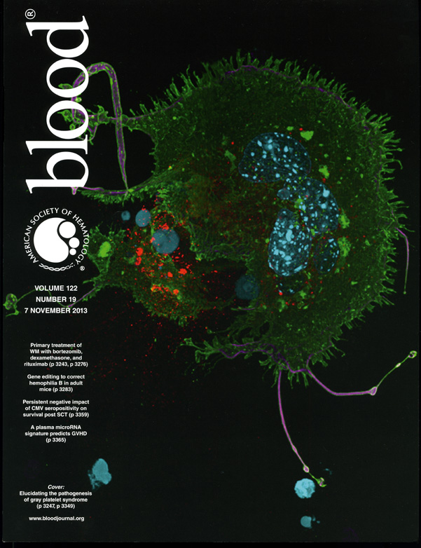
Blood, November 2013, an NBEAL2-knockout mouse megakaryocyte:
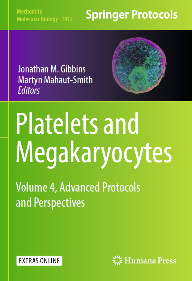
Cover of the recent Platelets and Megakaryocytes volume of Methods in Molecular Biology,
where we have a chapter on advanced fluorescence microscopy imaging methods:
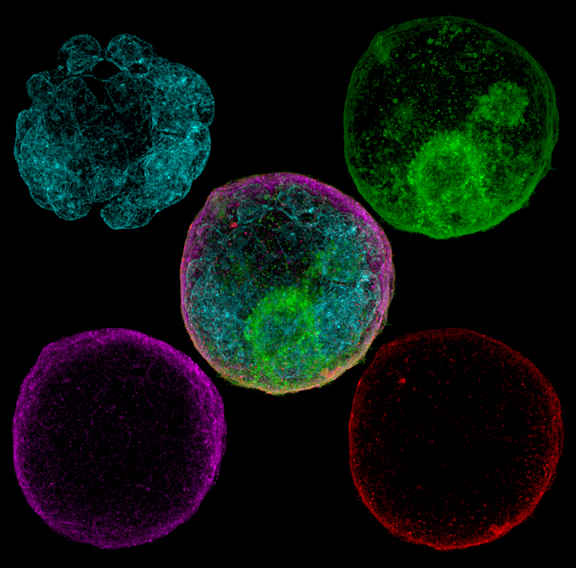
A better look at that cover image, deconstructed:
Blood, July 2015, showing a 3D render of a megakaryocyte:
ATVB, October 2018, showing a megakaryocyte taking up green fluorescent fibrinogen:

Winning image on the cover of Platelets for 2018, showing a proplatelet-forming megakaryocyte:

Molecular Cancer Research, November 2019, showing platelets adhering to cancer cells.

Blood, August 6 2020, showing NBEAL2 and SEC22B in a megakaryocyte.

Other images:
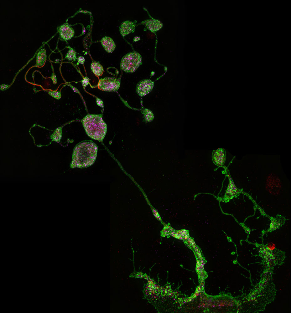
When megakaryocytes reach the final stage of maturity, they extend long proplatelets into the bloodstream that contain microtubules (magenta) that give rise to the cytoskeletal rings of platelets.
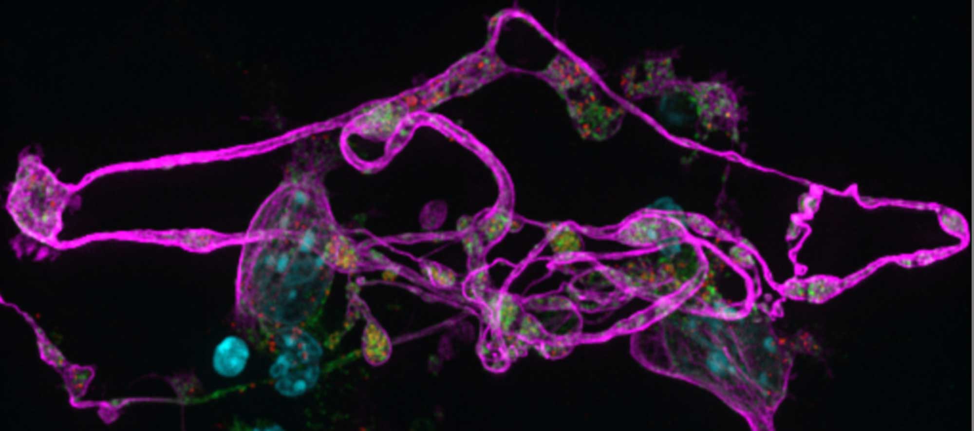
Multipanel SIM image of a platelet-forming mouse megakaryocyte
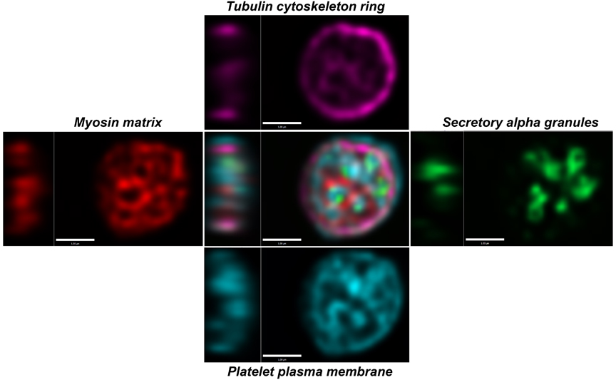
The end results are platelets containing structures that include a tubulin cytoskeletal ring, myosin matrix, secretory alpha granules and surface membrane:
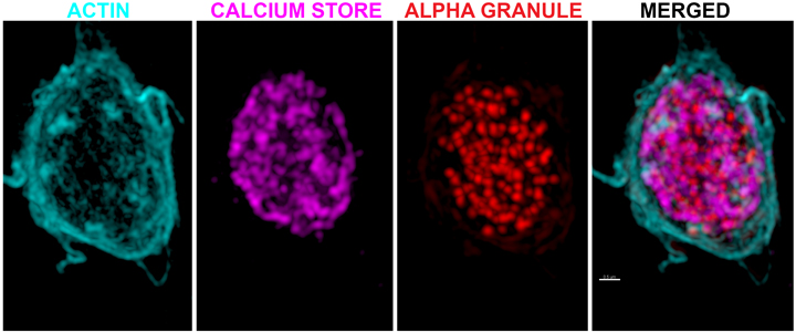
Platelets may be small, but they are complicated. Other aspects of their anatomy include an actin matrix, granule contents and a calcium store system: