Our research involves a wide range of reagents, methods, experimental approaches and technologies that are continually changing and advancing. Researchers at the SickKids RI are fortunate in having access to specialized facilities that provide advanced analytical capabilities, such as the SickKids proteomics, analytics, robotics and chemical biology centre (SPARC), and the Imaging Facility with state-of-the art optical and fluorescence microscopes. We also have access to a wide range of cutting-edge expertise and other support critical to research success. What follows are a few examples where access to world-class technical support has been critical to our research progress.


Genetic analysis used to take weeks or months and cost thousands of dollars, now it takes days or hours and costs much less. We have exploited ongoing advances in this field in several studies, including one that defined the genetic cause of a known hereditary condition: gray platelet syndrome (GPS), and one that discovered a new disease: ARPC1B deficiency.


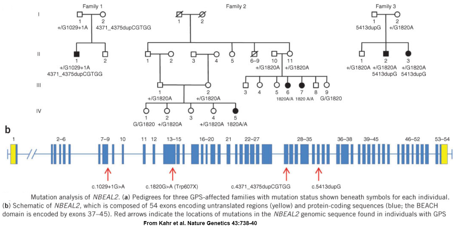
GPS was first reported in 1971, and a worldwide search for a cause was well underway when our relatively small international collaborative group became involved. We used homozygosity mapping of a chip-based genome-wide human single nucleotide polymorphism (SNP) array to localize the GPS locus to a 1.7 megabase region of chromosome 3 containing over 70 genes.  Sequencing all of them to find mutations would have taken a considerable effort, so instead we employed “next-generation sequencing” to analyzed mRNA from platelets – which may not have nuclei but they are produced by megakaryocytes which abundantly express RNA. We detected a transcript that was missing in GPS patients, and as a result we were one of 3 groups to simultaneously report loss of NBEAL2 expression as the molecular cause of GPS.


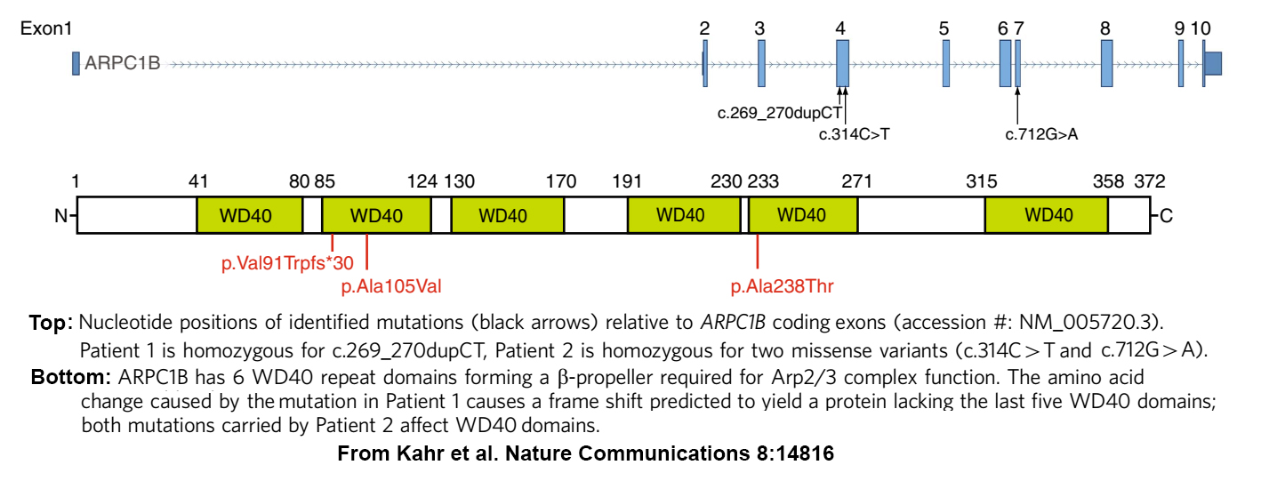
A few years later, we joined several SickKids colleagues in an effort to determine what was wrong with an especially challenging patient (see home page). At the time genomics analysis had reached the point where many hereditary disorders, including those affecting immunity/inflammation, platelets and the gastrointestinal system, had been linked to panels of genes where pathogenic and predisposing variants had been identified, or were predicted to occur. Unfortunately, scans of those panels for variants in our index patient consistently turned up negative. So eventually we used a less targeted approach and undertook a Whole Exome Sequence (WES) analysis of every gene expressed by the patient and his parents (and by another suspected patient and his parents). This search detected homozygosity for potentially pathogenic ARPC1B variants in both patients, for whom reduced or absent ARPC1B expression was confirmed by platelet protein analysis.


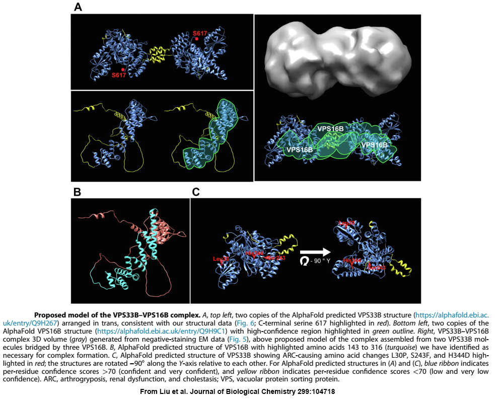
When we first linked VPS33B and VPS16B to platelet a-granule production and found these proteins formed a complex of unknown function, the obvious next step was to determine the structure of VPS16B-33B. At the time, the established way of doing this was to prepare milligram quantities of purified protein/complex, use it to form crystals, and perform X-ray diffraction analysis. Working with VPS16B-33B turned out to be challenging: we couldn’t make much of it, and it was not keen on forming crystals. Fortunately, other analytical options were available, including high-resolution cryo-electron microscopy (cryo-EM) established at the SickKids Nanoscale Biomedical Imaging Facility by our collaborator John Rubinstein. Using microgram amounts of protein we were able to use cryo-EM and other analytical techniques to determine the overall shape and size of the VPS16B-33B complex, and determine the number of components and their orientations. We also made use of recent advances in artificial intelligence by employing AlphaFold to obtain predicted protein structures that we were able to fit into the envelope of VPS16B-33B.


Each of us releases 100 billion platelets into our bloodstream every day. These tiny disk-shaped cells, only about 3 microns across, play vital roles as they patrol blood vessels for signs of injury. When they detect a breach they adhere, aggregate and activate formation of a clot that seals the wound and limits blood loss. Thus individuals lacking platelets or having dysfunctional cells experience abnormal bleeding. In addition to their key role in blood clotting, platelets are also involved in inflammation, immunity and wound healing, and in the formation of atherosclerotic plaques and pathological clots (i.e. thrombi) that trigger heart attacks and strokes. The many roles of platelets in health and disease are linked to their unique ability to store and release a wide range of biomolecules. These include a wide gamut of proteins stored within and released from secretory α‑granules, and studies of the development and function of these granules is a major focus of my research group.


High resolution laser fluorescence microscopy of a human platelet shows actin near the cell membrane and the internal calcium store system that surrounds abundant protein-carrying alpha granules.

Platelets are produced by megakaryocytes, cells that develop within the bone marrow until they reach enormous sizes of up to 100 microns across. At that point they generate extensions that protrude into blood vessels and shed platelets into circulation by the thousands.

Image of a cultured megakaryocyte (left, outer membrane in green) in the final stage of development when long extensions give rise to smaller anucleate bodies (right) that become platelets in vivo.

Many aspects of the cellular mechanisms involved in platelet production have remained obscure because megakaryocytes are difficult to study, and appropriate cell culture models were lacking until the advent of induced pluripotent cells. Platelets and megakaryocytes also present unique technical challenges, in that the former are tiny and abundant, while the latter are enormous and rare. We study megakaryocyte development in a variety of different ways using cells derived from humans and mice. During our studies we have employed, established and extended several specialized approaches, ranging from primary megakaryocyte culture to high resolution fluorescence and electron microscopy imaging. This has allowed us to study megakaryocyte and platelet structure/development in detail, and examine many novel aspects.

We have examined the passage of cargo proteins through vesicles of the endocytic pathway leading to α-granules, and observed how this traffic is perturbed in megakaryocytes lacking NBEAL2, or when vesicular pH is altered due to V-ATPase inhibition. We have also used high-resolution fluorescence imaging to identify NBEAL2 binding partners, map intracellular trafficking of thiol isomerases during development, examine the effects of expressing missense variants of ETV6, and count granules within individual megakaryocytes and platelets.

A major key to this success has been our access to advanced equipment, expertise and other critical support in state of the art facilities at the SickKids Research Institute, in particular the Imaging Facility.

Much of the recent progress that has been made in understanding platelet production, structure and function has come from studies of hereditary conditions where one or more of these aspects are affected. Patients with arthrogryposis, renal dysfunction and cholestasis (ARC) syndrome have platelets that lack α-granules. In a series of studies we identified that the root cause of this defect is loss of one of two proteins: VPS33B and VPS16B, which we showed using yeast two-hybrid screens and mass spectrometry form a functional protein complex. Ongoing studies are extending this work in several directions, including determining the structure of the functional VPS33B/VPS16B complex. This work recently culminated in the discovery that the VPS33B/VPS16B complex represents the first bidirectional SEC/MUNC complex with the potential to bind up to 4 SNAREs simultaneously (see Liu et al. J Biol Chem 2023 Jun;299(6):104718).

In another project we used classical genetics and platelet mRNA expression analysis to reveal the cause of gray platelet syndrome (GPS), where patients have platelets containing some alpha granule components, but normal granules fail to form. We found that GPS is caused by loss of function of NBEAL2 (neurobeachin-like 2), a large protein with several potential functional domains about which little is known. We have explored NBEAL2 function using Nbeal2-knockout mice that recapitulate many aspects of GPS pathology. These studies have allowed us to observe that loss of NBEAL2 function leads to impaired megakaryocyte development and affects their ability to package and retain protein cargo into alpha granules. Current studies are focused on elucidating the cellular mechanisms whereby NBEAL2 facilitates maturation and stability of alpha granules.

A recent example where the expertise of my group and our collaboration with others proved highly productive involved solving a mystery concerning a patient with a puzzling set of symptoms involving platelets and the immune system. We discovered the root of these problems was loss of expression of ARPC1B, a component of the Arp2/3 complex that generates branched actin filaments in blood cells. This work received considerable media interest and was recognized by the inaugural Janet Rossant Research innovation Prize, and it has stimulated further exploration of ARPC1B deficiency by us and others. A novel technical aspect of this project was the generation of gene knockout human megakaryocyte precursor (imMKCL) cells using CRISPR/Cas9 gene editing, which were used to model megakaryocyte development in the absence of ARPC1B function.

Fluorescence (left) and scanning EM (right) imaging of normal platelets spreading on fibrinogen (top) and platelets from a patient lacking ARPC1B (bottom) show a striking difference in the ability of the deficient cells to spread and form adherent lamellipodia via actin filament branching.


RESEARCH HIGHLIGHTS

Historical

Discovery of the genetic basis of Gray Platelet Syndrome. Preview

Identification of VPS16B, binding partner of VPS33B, as essential for alpha granule biogenesis. Read the paper.Accompanying editorial. 

Establishment of the Nbeal2 knockout mouse model of GPS – insights into its role in platelet development. Read the paper.Accompanying editorial.

Nature Genetics47,535–538 (2015):Germline mutations in ETV6 are associated with thrombocytopenia, red cell macrocytosis and predisposition to lymphoblastic leukemia.

FlnA binding to PACSIN2 F-BAR domain regulates membrane tubulation in megakaryocytes and platelets.Accompanying editorial.

 Inside Blood Editorial: Platelet Production – New Players in the Field

Arteriosclerosis Thrombosis and Vascular Biology 36:1164-73 (2016): Intracellular Trafficking, Localization and Mobilization of Platelet-Borne Thiol IsomerasesEditor’s choice free access.Accompanying editorial.

Blood Jun 9;127(23):2791-803 (2016) Plenary paper: A high-throughput sequencing test for diagnosing inherited bleeding, thrombotic, and platelet disordersAccompanying editorial.


Recent

Nature Communications 2017 April 3;8:4816: Loss of the Arp2/3 complex component ARPC1B causes platelet abnormalities and predisposes to inflammatory disease.

News Coverage: SickKidsCanadian Press CBC: WebsiteLead Story, The National April 3, 2017


Arteriosclerosis Thrombosis and Vascular Biology  https://doi.org/10.1161/ATVBAHA.118.311270 NBEAL2 (Neurobeachin-Like 2) Is Required for Retention of Cargo Proteins by α-Granules During Their Production by Megakaryocytes

Editorial: https://doi.org/10.1161/ATVBAHA.118.311614 Unlocking the Molecular Secrete(s) of α-Granule Biogenesis


Blood 2020 Aug 6;136(6):715-725. doi: 10.1182/blood.2019004276 The endoplasmic reticulum protein SEC22B interacts with NBEAL2 and is required for megakaryocyte α-granule biogenesis


Transfusion Medicine Reviews 2020 34:277-285 Inherited Platelet Disorders: Diagnosis and Management 


British Journal of Haematology 2022 197:245-246 Inherited platelet disorders: From new variants to new knowledge


Journal of Thrombosis and Haemostasis 2022 20:1712-1719 Platelet VPS16B is dependent on VPS33B expression, as determined in two siblings with arthrogryposis, renal dysfunction, and cholestasis syndrome


Platelets 2023 34:2157808 Evaluation of human platelet granules by structured illumination laser fluorescence microscopy 


Journal of Biological Chemistry 2023 299:104718 The Sec1-Munc18 protein VPS33B forms a uniquely bidirectional complex with VPS16B


Research and Practice in Thrombosis and Haemostasis 2024 8:102352 Human platelets contain a pool of free zinc in dense granules


Visit PubMed for a full list of publications.