Helicobacter pylori (H. pylori)

Helicobacter pylori (H. pylori) infects 50% of the world’s population and can lead to peptic ulcer disease and gastric cancer. In fact, H. pylori is the single most important risk factor for the development of gastric cancer. To cause a chronic infection, H. pylori must possess mechanisms that allow it to evade host responses. The Jones laboratory is focused on determining these mechanisms and how they may be related to disease in particular gastric carcinogenesis.

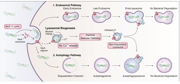
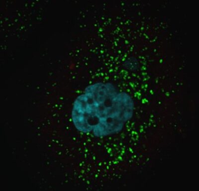
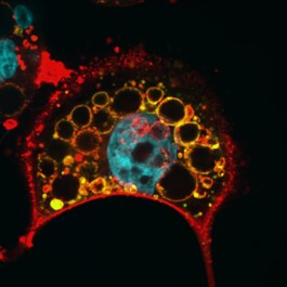
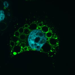
We are currently investigating how the Vacuolating cytotoxin (VacA), a virulence factor secreted by H. pylori, alters host cell trafficking to develop an intracellular niche for the bacteria where it can survive in parietal cells and escape from antibiotic eradication therapy. We have determined that VacA inhibits a lysosomal calcium channel TRPML1 to create this niche. Furthermore, activating TRPML1 eliminates the niche and kills the bacteria. We are now interested in targeting this pathway as a potential novel therapeutic. We are also interested in how inhibition of this channel can alter parietal cell function to promote disease such as gastric cancer.

An illustration of the mechanism by which the VacA toxin secreted by H. pylori inhibits the lysosomal Ca2+ channel TRPML1 and impairs the vesicular trafficking leading to non-degradative lysosomes. These dysfunctional lysosomes are not capable of killing the intracellular H. pylori.
A microscopic image of a cell stained for a lysosomal marker, where the healthy lysosomes within the cell are seen as hundreds of small green dots.
A microscopic image of a VacA-treated cell stained for both, a lysosomal marker in green and VacA in red. The enlarged dysfunctional lysosomes are seen as medium-sized yellow circles due to the co-localization of both markers. VacA red staining also delineates the plasma membrane
A microscopic image of a VacA-treated cell stained for a lysosomal marker. The enlarged dysfunctional lysosomes or vacuoles are seen as about 20 medium-sized green circles.
A microscopic image of a VacA-treated cell stained for VacA in red. VacA red staining delineates the enlarged dysfunctional lysosomes and the plasma membrane of a cell .

H. pylori VacA toxin transforms normal lysosomes (labelled by Lamp1 in green) into huge vacuoles to promote intracellular bacterial survival

A microscopic image of purple-stained gastric epithelium from a patient infected with a VacA+ H. pylori. Small clusters of red dots represent intracellular H. pylori bacteria within gastric cells
A high magnification microscopic image of purple-stained gastric epithelium from a patient infected with a VacA+ H. pylori. H. pylori staining in red show large clusters of intracellular bacteria in a parietal cell.

H. pylori staining (red) in human gastric biopsies

Inflammatory Bowel Disease (IBD)

Canada has one of the highest rates of inflammatory bowel disease in the world. Our research team is interested in understanding how host genetic risk factors and environmental factors associated with IBD promote disease in the hopes of developing novel solutions to treat or prevent disease.

We are interested in two main genetic risk factors, ATG16L1 and Nod2 and how they influence autophagy, the cells garbage disposal system to cause disease.

We are also interested in vitamin D, as vitamin D deficiency is linked to IBD and more severe disease. We have shown that vitamin D deficiency, an environmental risk factor that is associated with IBD alters microRNA that target autophagy genes, including ATG16L1 in the intestine of mice and in involved tissue from IBD patients. We are interested in the effect of vitamin D deficiency in the context of IBD genetic risk factors.

A Venn-diagram explaining how genetics, environmental factors, microbiome, and immune system intersects in the pathogenesis of IBD. The Jones lab studies on genetic factors like Nod2 frame shift and ATG16L1, and environmental factors like Vitamin D deficiency.
A microscopic image of small intestines obtained from control and vitamin D-deficient mice stained in red for a marker of Paneth cell granules. On the left, in the control small intestine, the red clusters of Paneth cell granules are homogenous in size and number. On the right, red clusters of Paneth cell granules are larger or diffuse in the vitamin D-deficient mouse.

Vitamin D deficient (VDD) mice have abnormal Paneth cell granules (UEA-1 staining)