MINFLUX is a method of imaging that is able to synergistically combine the strengths of STED and PALM/STORM to achieve an unprecedented 3D localization precision of 1-3 nm, and can also be applied to the recording of molecular trajectories with frequencies of up to 10 kHz. In MINFLUX imaging, fluorophores are individually switched in a manner similar to SMLM, allowing for the separation of overlapping signal at a single-molecule level. In turn, the fluorophore localization is accomplished using a movable structured excitation beam featuring a central intensity minimum (zero). Localization is performed by actively targeting the zero of the excitation donut. As the intensity minimum of the excitation beam approaches the fluorophore, there is a corresponding decrease in the emitted fluorescence per unit excitation power, thereby indirectly revealing the residual distance of the molecule to the excitation zero. Through a series of calculated iterations, the excitation minimum is brought as close as possible to the molecule until the detected fluorescence rate matches that of the background noise. At this point, the perfectly controlled position of the zero-excitation intensity becomes a proxy for the position of the emitter.
SUPER RESOLUTION
MINFLUX
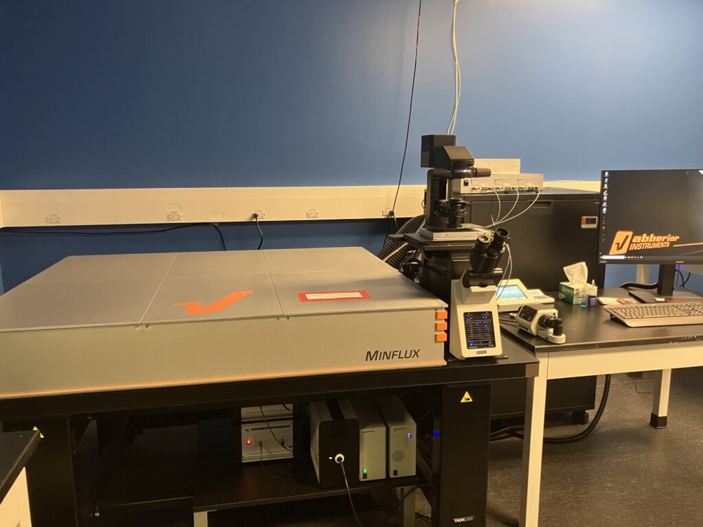
System name: Abberior MINFLUX
Objectives
- 100x/1.45 (Minflux, confocal)
- 40x/0.95 (Widefield, eyepiece)
- 20x/0.8 (Widefield, eyepiece)
Detectors
- 5x APD: DAPI (422nm – 467nm); GFP (500nm – 550nm); Cy3 (580nm – 630nm); Cy5 Near (650nm – 685nm); Cy5 Far (685nm – 720nm); PMT (Alignment)
Lasers
- Minflux – 640 nm
- Confocal – 561 nm, 488 nm
- Activation – 405 nm
Acquisition Modes
- 2D
- 3D
- Fast 2D tracking
- 3D tracking
Structured Illumination Microscopy (SIM)
SIM is an imaging technique that employs patterned excitation light to generate interference patterns (moiré patterns) that contain high frequency spatial information. By collecting a series of images with different interference patterns, specialized algorithms are able to extract super-resolution detail to produce a reconstructed final image that has approximately twice the resolution of traditional diffraction limited microscopes, ie. approx. 110 nm (XY), 300 nm (Z). This technique is ideally suited for 3D imaging of thin (<20 um), fixed samples that have excellent fluorescence labeling.
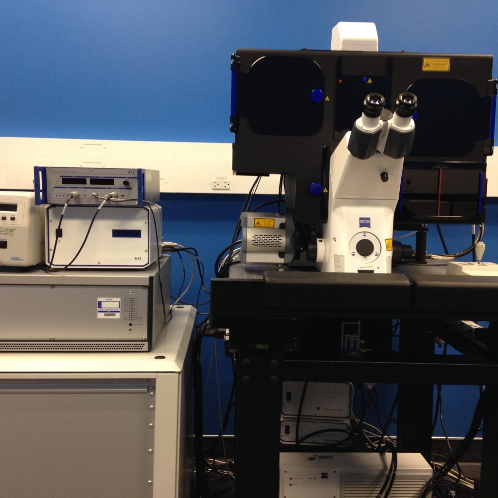
System name: Zeiss Elyra PS1
Objectives
- 63x/1.4 PlanApo
- 100x/1.46 alpha PlanApo
Camera
- Andor iXon3 885 (SIM)
Lasers
- 405 nm (150 mW)
- 488 nm (200 mW)
- 561 nm (200 mW)
- 640 nm (150 mW)
Localization Microscopy (STORM/PALM)
Stochastic optical reconstruction microscopy (STORM) and photoactivated localization microscopy (PALM) function by stochastically cycling fluorophores between ‘on’ and ‘off’ states in a controlled manner, followed by precise localization of the centre of each fluorescent molecule to within tens of nanometers. By processing thousands of images, the molecular coordinates of all labeled molecules are obtained with a resolution ten times better than traditional diffraction limited microscopes, i.e. approximately 20 nm (XY), 50 nm (Z). This technique is best suited for imaging the stoichiometric architecture of proteins within defined complexes, and is largely limited to fixed, cellular samples.

System Name: Zeiss Elyra PS1
Objectives
- 63x/1.4 PlanApo
- 100x/1.46 alpha PlanApo
Cameras
- Andor iXon DU897 (PALM)
Lasers
- 405 nm (150 mW)
- 488 nm (200 mW)
- 561 nm (200 mW)
- 640 nm (150 mW)
Other
- 3D: double-helix PSF
Stimulated Emission Depletion (STED)
STED microscopy overcomes the diffraction limit by overlapping the primary excitation laser with a secondary, donut-shaped, high-power laser beam. Irradiation by the secondary laser forces molecules in the outer region of the primary excitation area to return to the ground state, while leaving fluorophores in the centre of the excitation focus to form the high resolution image. This application can achieve resolutions up to 50 nm laterally, and up to 120 nm axially, and is best suited for 2D or 3D imaging of thin samples (<50 um) that have excellent fluorescence labeling.
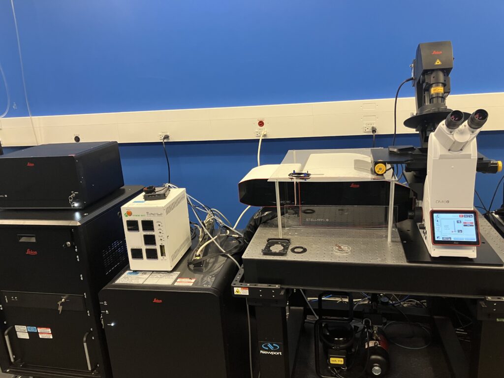
System Name: Leica Stellaris 8 FALCON w/STED
Objectives
- 93x/1.3 STED (motCORR)
- 100x/1.4 STED
Lasers
- 405 nm
- white light laser (pulsed, 440-790 nm)
- 775 nm (pulsed, STED)
Other
- TauSTED
CONFOCAL
Confocal microscopy is a powerful technique in which a pinhole is used to block out-of-focus light in image formation, thereby generating improved contrast and resolution compared to widefield microscopes. Generally speaking, there are two types of confocal microscopes: point scanning and spinning disk. Although slower than a spinning disk confocal, point scanning confocal microscopes tend to achieve higher resolutions, and have more integrated solutions for specialized techniques such as fluorescence recovery after photobleaching (FRAP). On the other hand, spinning disk confocals tend to have significant advantages in both speed and phototoxicity, making them ideally suited for live cell imaging.
In recent years, the resolution of traditional point scanning confocal microscopes has been extended to the super-resolution realm by implementing three key components to a workflow:
- Closing the pinhole diameter below 1 Airy Unit,
- Using low-noise, high-sensitivity detectors,
- Combining pixel oversampling with modern deconvolution algorithms.
Taken together, these implementations can improve resolution by up to 40% compared to traditional confocal microscopes, i.e. 120 nm (XY), 400 nm (Z), and can be applied to most standard confocal applications.
A specialized application of confocal microscopy is multiphoton imaging. In this technique, a specialized infrared laser is used to selectively excite molecules located within a femtoliter volume of space, up to one millimetre deep within a sample. The primary advantage of multi photon imaging is reduced phytotoxicity and deep sample penetration, making it ideal for intravital microscopy.
Point scanning confocals
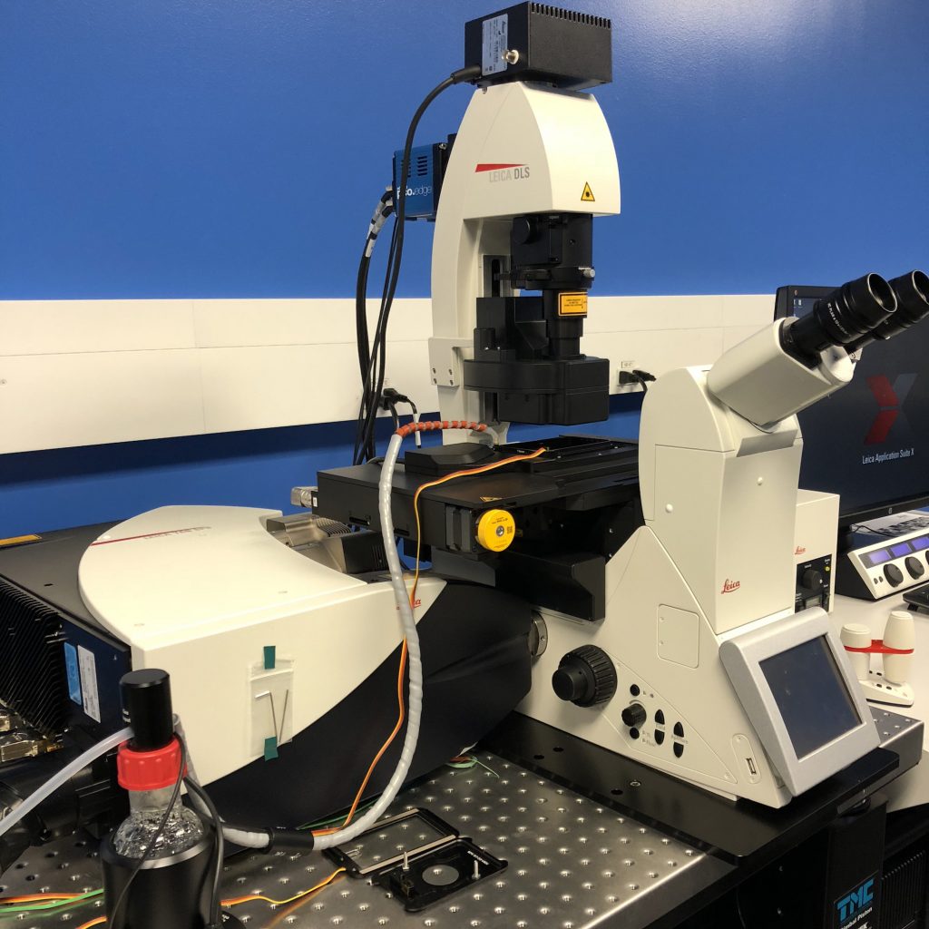
System Name: Leica SP8 Lightning Confocal/Light Sheet
Objectives
- 5x/0.15
- 20x/0.75
- 40x/1.3 (motCORR)
- 63x/1.3
Lasers
- 405 nm
- 448 nm
- 488 nm
- 552 nm
- 638 nm
Detectors
- 4x HyD, 1x PMT, resonance scanner
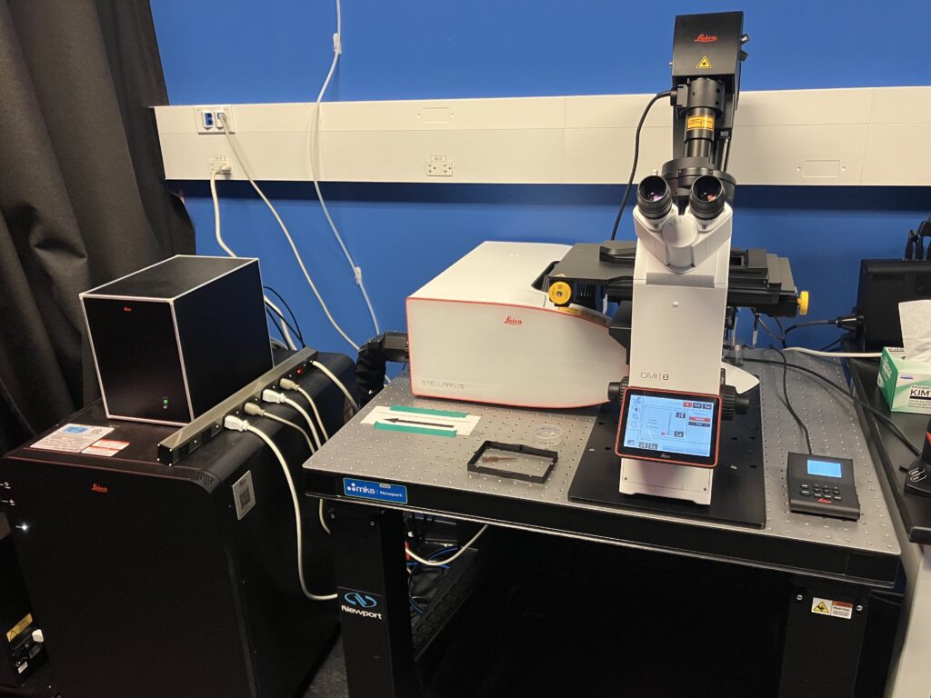
System name: Leica Stellaris 5 WLL
Objectives
- 5x/0.15
- 10x/0.4
- 20x/0.75
- 40x/1.1
- 63x/1.4
Lasers
- 405 nm
- white light laser (pulsed 485-685 nm)
Detectors
- 4x HyD S
Other
- Resonance scanner (2048 x 2048)
- Lightning Deconvolution
- TauContrast

System name: Leica Stellaris 8 FALCON w/STED
Objectives
- 10x/0.4
- 20x/0.75
- 40x/1.3
- 63x/1.4
Lasers
- 405 nm
- white light laser (pulsed 440-790 nm)
Detectors
- 2x HyD S, 2x HyD X, HyD R
Other
- Lightning Deconvolution
- TauContrast
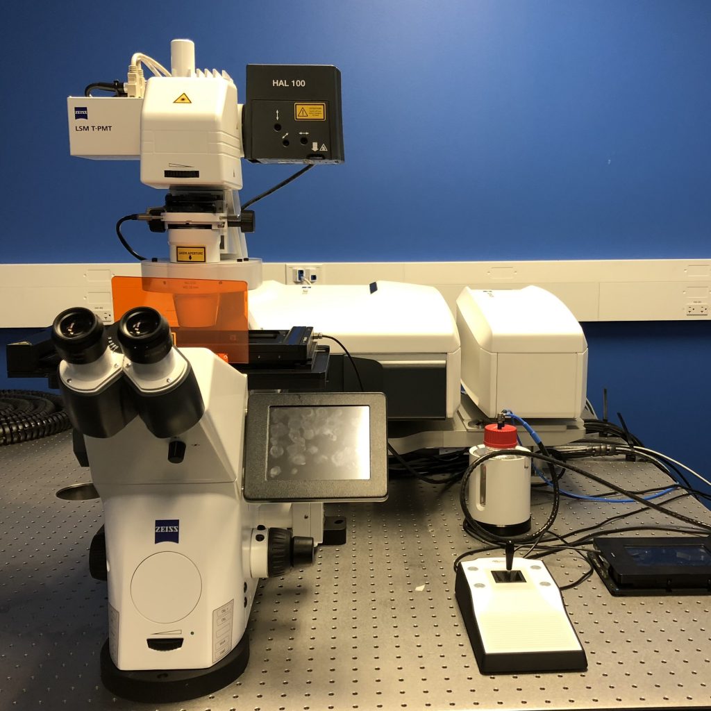
System Name: Zeiss LSM880 Airyscan
Objectives
- 5x/0.16
- 10x/0.45
- 20x/0.8 PlanApo
- 40x/1.1
- 63x/1.4 PlanApo
Lasers
- 405 nm
- argon (458/476/488/496/514 nm)
- 552 nm
- 642 nm
Other
- 32-channel spectral PMT, Airyscan Fast
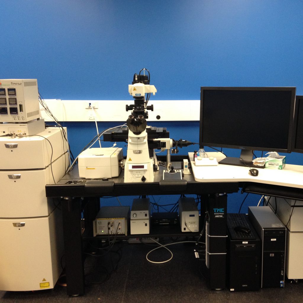
System Name: Nikon A1R Confocal Microscope w/FCS + FLIM
Objectives
- 10x/0.5
- 20x/0.75
- 40x/1.25
- 60x/1.4
- 100x/1.4
Lasers
- 402 nm
- 440 nm (pulsed)
- 488 nm
- 561 nm
Detectors
- 2 x GaAsP PMT, 2 x PMT
- 32-channel spectral PMT
Other
- HD resonance scanner (1024 x 1024)
- Denoise.ai, NIS-Elements Deconvolution
Spinning disk confocals
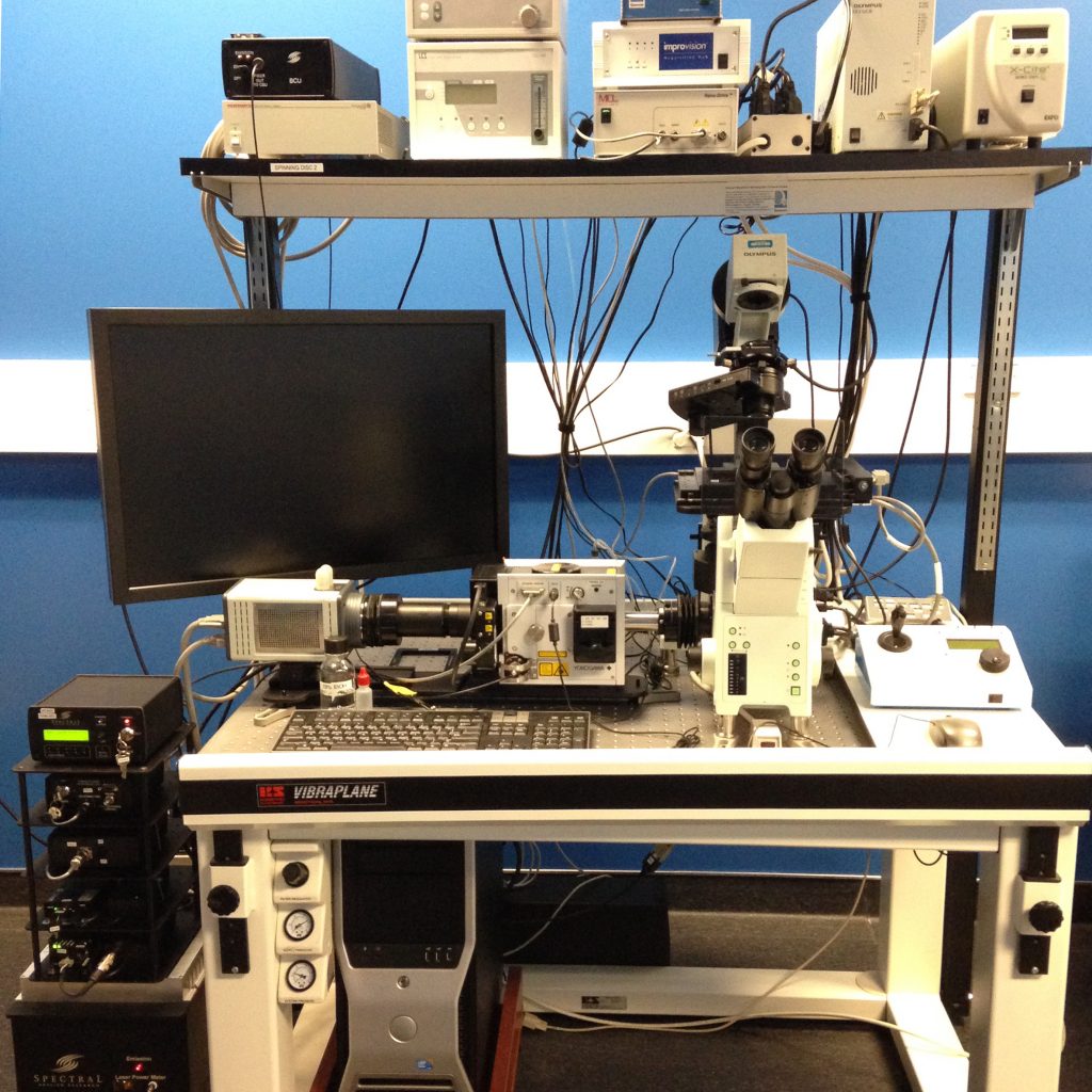
System Name: Quorum Spinning Disk 2
Objectives
- 10x/0.4
- 20x/0.75
- 40x/0.9
- 60x/1.27
- 60x/1.35
Lasers
- 405 nm
- 491 nm
- 561 nm
- 642 nm
Camera
- Hamamatsu C9100-13 EM-CCD
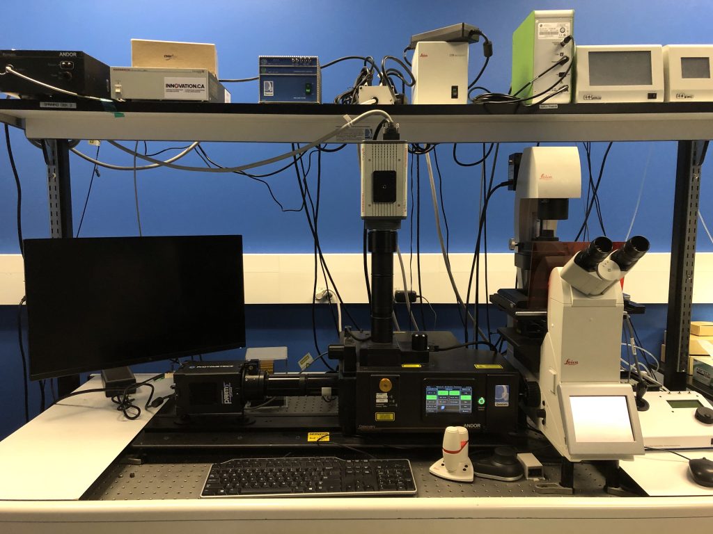
System Name: Quorum Spinning Disk 3
Objectives
- 10x/0.4
- 20x/0.8
- 40x/1.1
- 40x/1.3
- 63x/1.4
Lasers
- 405 nm
- 491 nm
- 561 nm
- 637 nm
Camera
- Hamamatsu C9100-13 EM-CCD, Photometrics Prime 95B
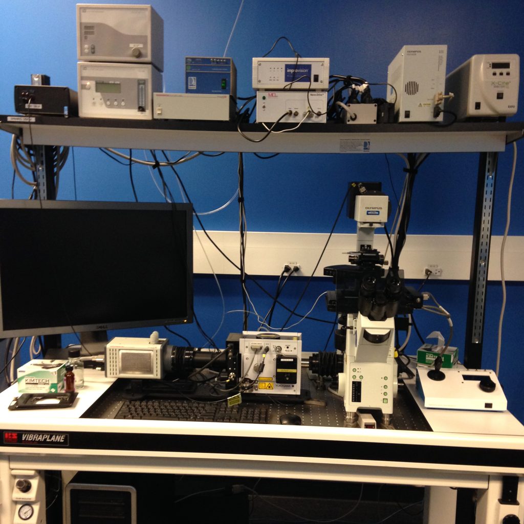
System Name: Quorum Spinning Disk 4
Objectives
- 4x/0.16
- 10x/0.4
- 20x/0.75
- 40x/0.95
- 60x/1.35
- 100x/1.4
Lasers
- 405 nm
- 491 nm
- 561 nm
- 642 nm
Camera
- Hamamatsu C9100-13 EM-CCD
Multiphoton imaging
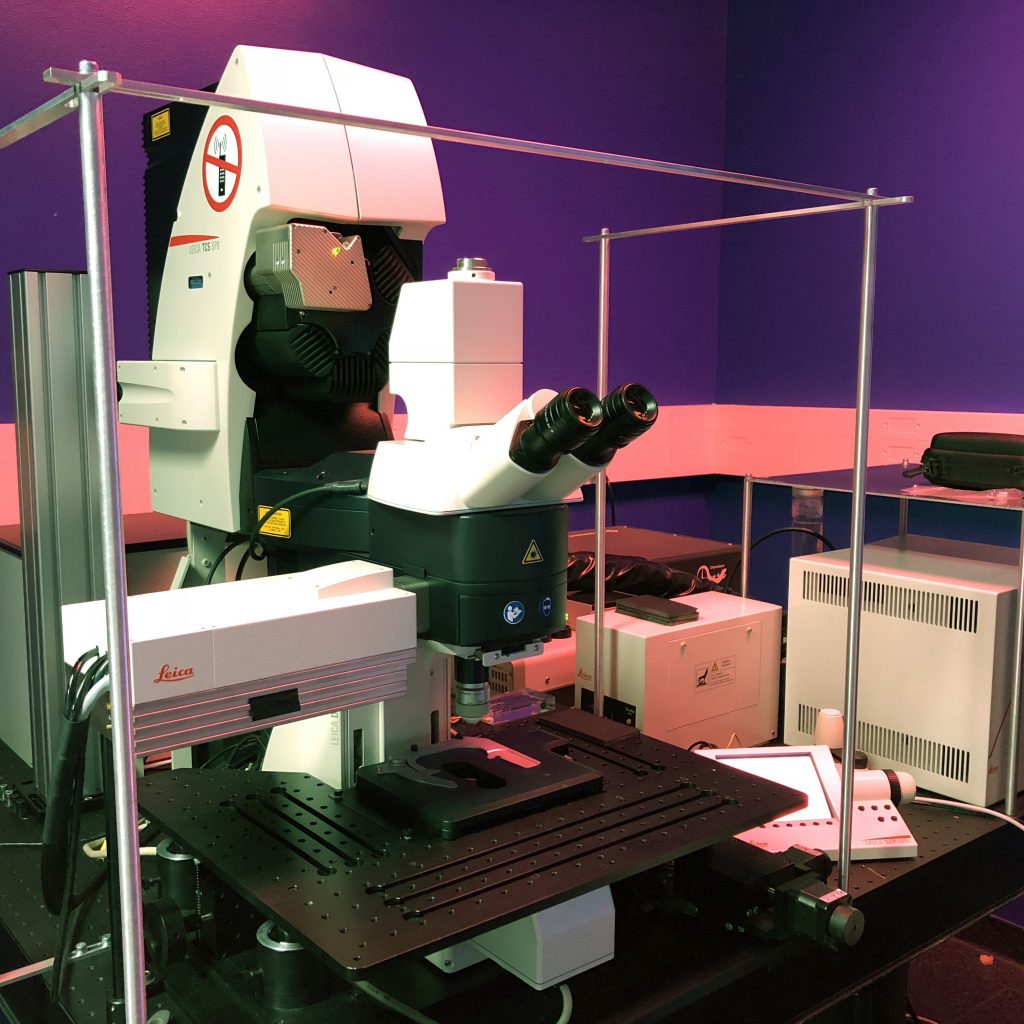
System Name: Leica SP8 Confocal w/Two-Photon
Objective
- 25x/0.95
Detectors
- 1 x HyD, 2 x PMT, 2 x NDD-HyD
Lasers
- Argon (458/476/488/496/515 nm)
- 561 nm
- 633 nm
- Coherent Ultra II (680-1080 nm, >3.3 W)
WIDEFIELD
Widefield imaging is the method of choice for basic fluorescence imaging. It is most often applied to examining protein expression levels in cells and tissue, general patterns of localization, and population studies. It is especially powerful for high-speed imaging, as well as ratiometric measurements of intracellular pH, or other ion levels. Finally, it is the tool of choice for researchers looking to image cellular samples with white light (eg. DIC imaging of wound assays, beating cilia, etc.).
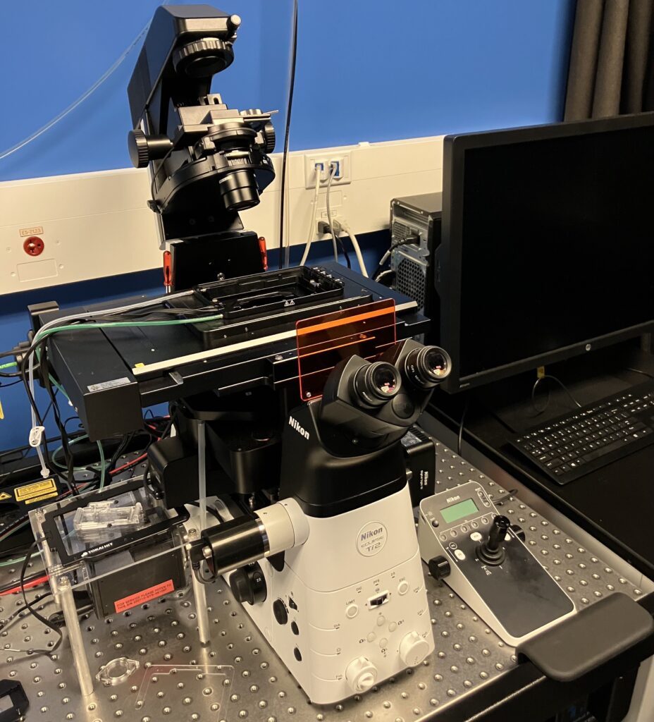
System Name: Nikon Epifluorescence Microscope w/TIRF
Objectives
- 4x/0.2
- 10x/0.45
- 20x/0.8
- 40x/1.25 (Sil)
- 40x/1.3 Super Fluor (O)
- 60x/1.4 (O)
- 60x/1.49 (O) TIRF
Camera
- Hamamatsu Orca Fusion BT (Fluorescence)
- Nikon DS10 (Colour)
Light Source
- Spectra III (Excitation: 348 nm, 380 nm, 434 nm, 475 nm, 511 nm, 555 nm, 637 nm, 748 nm)
- Lasers (TIRF): 488 nm (90 mW), 561 nm (70 mW)
Filter Cubes
- Fura2/TRITC, Penta (390/475/555/635/747), Triple (440/510/575), TIRF (488/561)
- Emission Filters: 432/36, 474/27, 515/30, 544/24, 595/31, 680/42, TIRF 525/50, TIRF 600/50
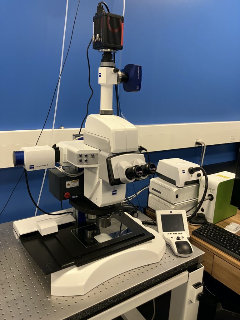
System Name: Zeiss AxioZoom V16
Objectives
- 1.0x/0.25
- 2.3x/0.57
Zoom
- 0.7x – 11.2x
Camera
- pco.edge 4.2 bi (Fluorescent)
- Axiocam 305 color (Colour)
Filter Cubes
- DAPI
- GFP
- RFP
- Cy5
OTHER
Light sheet fluorescence microscopy
In light sheet imaging, samples are illuminated perpendicularly to the direction of observation using a thin sheet of light that is projected through the sample. Although the lateral resolution of a light sheet microscope is comparable to a widefield microscope, its key advantages are greatly reduced phototoxicity, rapid imaging, and the ability to image large samples from multiple directions for increased clarity and resolution. This technique has been masterfully adapted for use with cleared tissues, such as brain, kidney, and other organs measuring up to several millimetres in thickness
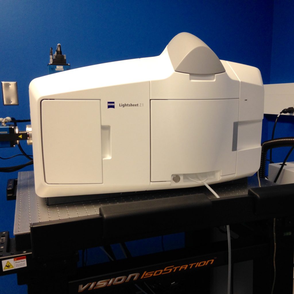
System Name: Zeiss Light Sheet Z1
Illumination Objectives
- 5x/0.1
- 10x/0.2
Detection Objectives
- 5x/0.16
- 10x/0.5
- 20x/1.0
- 20x/1.0 (CLARITY, RI=1.45)
Lasers
- 405 nm
- 488 nm
- 561 nm
- 638 nm
Cameras
pCO Edge 5.5 x 2
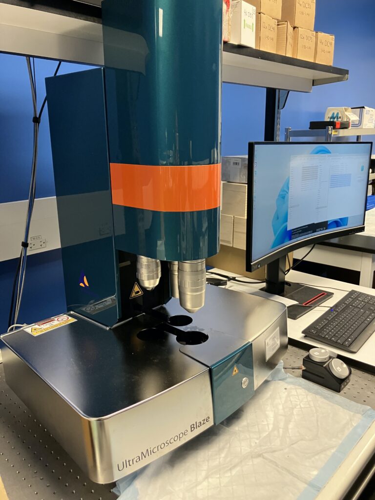
System Name: Miltenyi Biotec UM Blaze
Objectives
- 1.1x/0.1
- 4.0x/0.35
- 12x/0.53
Dipping Caps
- 1.1x DC40 (RI 1.33-1.49)
- 1.1x DC57 (RI 1.50-1.57)
- 4.0x DC33 (RI 1.33 – 1.41)
- 4.0x DC49 (RI 1.42-1.48)
- 4.0x DC57 (RI 1.49-1.57)
- 12x DC33 (RI 1.33-1.41)
- 12x DC49 (RI 1.42-1.48)
- 12x DC57 (RI 1.49-1.57)
Lasers
- 405 nm
- 488 nm
- 561 nm
- 639 nm
- 785 nm
Cameras
pco.edge 4.2
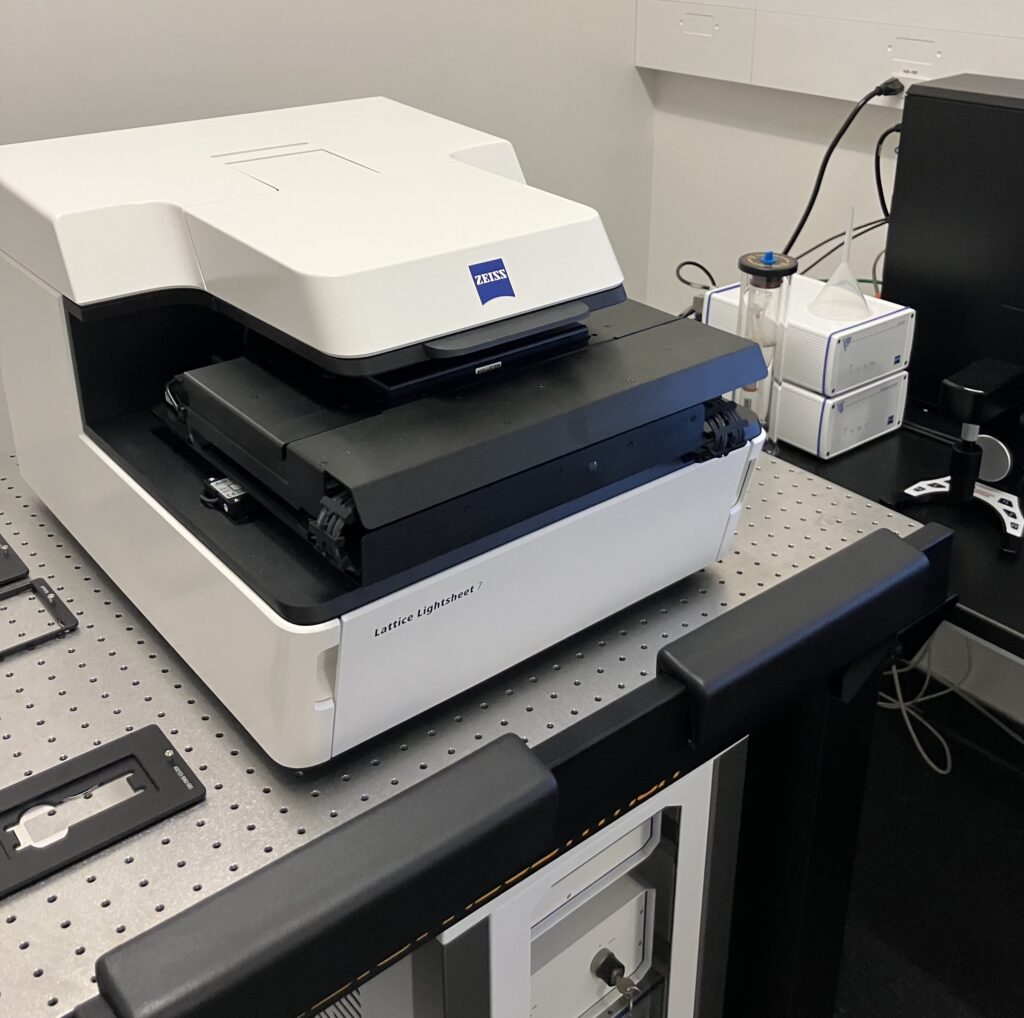
System Name: Zeiss Lattice Light Sheet Microscope
Objectives
- llumination Lens: 13.3x/0.4 (30 degrees)
- Detection Lens: 44.8x/1.0 (60 degrees)
Camera
- pco.edge 4.2
Lasers
- 488 nm
- 561 nm
- 640 nm

System Name: Leica SP8 Lightning Confocal/Light Sheet
Illumination Objectives
- 6x/0.05
- 5x/0.07
- 5x/0.15
Detection Objectives
- 5x/0.15
- 10x/0.3
- 25x/0.95
TwinFlect Mirrors
- 5 mm
- 5 mm
- 8 mm
- 8 mm (glycerol)
Lasers
- 405 nm
- 448 nm
- 488 nm
- 552 nm
- 638 nm
Detectors
- pCO Edge 5
Fluorescence Lifetime Imaging (FLIM)
FLIM is an excellent technique for mapping the local environment and molecular interactions of a fluorophore. A fluorophore’s lifetime (ie. the amount of time spent in the excited state) is a quantitative signature that can be used to extract information about its surrounding environment: ion concentrations, pH, viscosity, and molecular interactions. Furthermore, because fluorescence lifetime measurements are not affected by probe concentrations or photobleaching, and are independent of excitation, FLIM is an ideal tool for measuring FRET in living cells.

System Name: Leica Stellaris 8 FALCON w/STED
Objectives
- 20x/0.8
- 40x/1.3
- 60x/1.4
- 93x/1.3
- 100x/1.4
Lasers
- white light laser (pulsed, 440-790 nm)
Detectors
- 2x HyD S, 2x HyD X, HyD R
Other
- Leica FALCON FLIM module
Fluorescence Correlation Spectroscopy (FCS)
FCS is a correlative analysis of fluorescence fluctuations over time within a defined volume. As fluorophores enter and exit the observed volume, the resulting fluctuations in intensity encode parameters such as diffusion rates, molecular volume, binding affinities, and colocalization. This tool is especially useful for measuring the mobility of rapidly diffusing cytosolic proteins.

System Name: Leica Stellaris 8 FALCON w/STED
Objectives
- 40x/1.1
Lasers
- white light laser (pulsed, 440-790 nm)
Detectors
- 2x HyD S, 2x HyD X, HyD R
Other
- Leica FALCON FCS module
Total internal reflection fluorescence (TIRF)
TIRF microscopy is a specialized technique that allows for imaging of fluorescent molecules located within 200 nm of the coverslip. It accomplishes this by the generation of a fluorescent evanescent wave that decays exponentially as it projects upward from the surface of the coverslip. In doing so, only fluorophores located 100-200 nm from the coverslip are excited, resulting in very high signal-to-noise imaging of the basolateral surface of cells. This technique is especially useful for observing membrane associated processes such as ligand binding, cell adhesion, and vesicle trafficking from the cell surface.
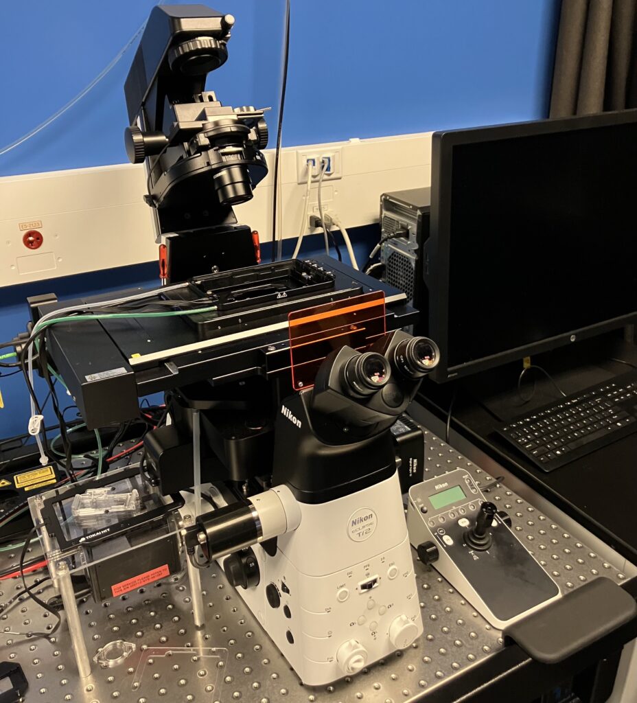
System Name: Nikon Epifluorescence Microscope w/ TIRF
Objective
- 60x/1.49 (O)
Camera
- Hamamatsu ORCA-Fusion BT
Lasers
- 491 nm
- 561 nm
Automated slide scanning
Digital slide scanning is the ideal solution for imaging whole tissue sections. Up to 250 slides can be automatically scanned in a high-throughput fashion using high-resolution 20x and 40x objectives, in both brightfield and fluorescence modes. The resulting images can be analyzed to provide area quantification, complex colour analysis, cytonuclear quantitation, etc. This technology is featured as a service in the Facility, with the turnaround typically being 24 hours.
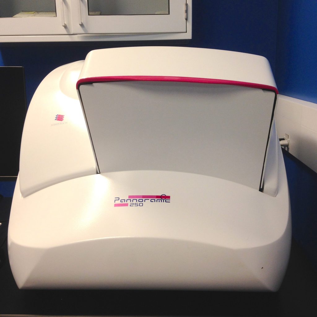
System Name: 3DHistech Pannoramic Flash II Slide Scanner
Objectives
- 20x/0.8
- 40x/0.95
Cameras
- pCO Edge 4.2 sCMOS
- CIS VCC-FC60FR19CL
Fluororescence Filter Cubes
- DAPI
- GFP
- Cy3
- Cy5
- Quad cube
All Imaging Facility equipment can be booked through QReserve.
