Ectodermal remodelling

Ectoderm is an epithelial layer that becomes distinct during gastrulation.  Early embryonic appendages and organ primordia are commonly surrounded by a single cell layer of ectodermal cells.  Interestingly, cells within these sheets move about and exchange places with their neighbours in 2D.

Cell rearrangements accomodate rapid addition of new cells and control the shape of ectodermal tissue over time. These rearrangements are the combined result of cell-intrinsic and -extrinsic forces.  Individual cells control how tightly they adhere to their neighbours at junctions and how they contract actomyosin networks that affect cell shapes and movements.  The behaviours of individual cells influences tissue forces that then feed back upon cells to modify their movements.  Our aim is to define how specific pathways influence the physical properties of individual cells and the transduction of force between cells.

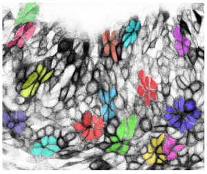
Ectoderm in the flank and early limb field remodels through cell neighbour exchanges.
A multicelluar rosette resolves to form two rows of cells along the dominant axis of tissue tension.
Rosette formation during ectodermal remodelling

Small groups of cells coordinate their behaviours to shape a tissue sheet.  These behaviours are regulated by cell-intrinsic and -extrinsic forces.

Mesodermal morphogenesis

Multiple growing structures in the embryo such as the limb buds and branchial arches are composed of a mesodermal core surrounded by ectoderm.  Although our field has made substantial progress in understanding morphogenesis of a tissue sheet in 2D, less is known about how a volume of tissue achieves shape change over time.

Unlike mesenchyme in mature tissues, mesodermal cells in the embryo are often intimate neighbours without much intercellular matrix.  Based on analogy to epithelia, we presume that mesodermal cells negotiate their positions among their neighbours in 3D.  In our lab, we study those cell behaviours using a combination of mouse genetics, live imaging, physical assessments and mathematical modelling.  Our goal is to achieve an integrated view of how signalling pathways modulate the physical environment to influence cell behaviours and shape tissue.

3D shape of a developing limb bud

Formation of the nuanced 3D shape of the limb bud precedes skeletal pattern formation.  Mutations that cause profound congenital limb deficiency often alter this shape.

Mesodermal cells move in an orderly fashion into the early limb field (nuclei are visualised).
Tracking of branchial arch cell movements in 4D.
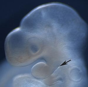
Morphology of a branchial arch in a developing fetus

The first branchial arch has a distinct 3D morphology.  We aim to understand how that morphology is generated.

Skeletal pattern formation

Biochemical signals may specify pattern by influencing downstream molecular networks and by shaping tissue.  We examine transcriptional and nontranscriptional means by which Iroquois homeodomain proteins Irx3 and Irx5 influence skeletal pattern in the limb.  Interestingly, although Irx3/5 are required to counterbalance Sonic hedgehog (Shh) to generate a full complement of skeletal elements, they are not required to specify pattern per se since pattern is restored by depleting all three genes.  Currently, we are exploring a morphogenetic role for Irx3/5 that shapes the limb field in advance of pattern formation.

Skeletal patterning of the hind limb

Hindlimb skeletal pattern requires balance between Irx3/5 and Shh gene functions.