X-Ray Computed Tomography (Micro CT)
The micro CT facility provides access to powerful sub-micron X-ray microscope (XRM) that delivers spatial resolution down to ~600 nm (with voxel sizes as small as 70 nm) while preserving a large field of view. It excels at non-destructive 3D histology, in-situ or hydrated imaging, and correlative workflows that complements downstream FIB-SEM, TEM, or light microscopy. Our team is available to assist with sample preparation, scan design, contrast optimization, and data integration into your imaging pipeline.
Equipment
Zeiss Versa 510 XRM
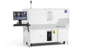
The Zeiss Xradia 510 Versa is a 3D X-ray microscope engineered for high-resolution, non-destructive imaging of a wide range of samples. It delivers advanced micro-computed tomography (micro-CT) with exceptional performance, achieving true spatial resolution down to 600 nm and voxel sizes as small as 70 nm, making it ideal for detailed internal visualization across materials and life sciences.
Why X-ray microscopy ?
• Non-destructive 3D imaging
• Image live or hydrated biological specimens
• Enable correlative workflows with FIB-SEM and light microscopy
• Perform virtual histology on intact samples
• Visualize cellular and subcellular architecture
• Generate virtual cross-sections at any depth
• Assess microstructure prior to ultramicrotomy
• Examine contrast-enhanced soft tissues
• Image entire small organisms or organs
Features
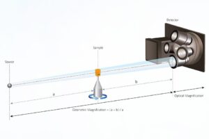
Powered by dual-stage magnification, the Versa XRM leverages advanced synchrotron-grade optics and proprietary RaaD (Resolution at a Distance) technology to deliver high-resolution imaging—even at extended working distances.
• 10 Watt Power
• Source Voltage 30 to 160kV
• Objective lenses: 0.4x , 4x , 20x , 40x
• X-ray filters: 6 LE and 6 HE (12 total)
• 4MP CCD camera
• 360 stage rotation at 0.005 degree accuracy
Services
X-Ray Computed Tomography
We will help designing the scan and parameters to achieve the best tomogram. Fast scans can be done in one hour while high resolution scans require longer acquisition time and more accurate slices.
Correlative Microscopy
It can be challenging to determine where a specific cell or structure is located inside a larger organ or tissue. Correlative X Ray-EM (XREM) imaging is a technique to assist down stream imaging. XREM merges the power of rapid non-destructive x-ray microscopy with the high resolving power of EM. We use a suite of software such a Dragonfly or Amara to correlate the data. The technique can be used for super resolution light microscopy as well as FIB-SEM, array tomography and TEM imaging.
Sample Preparation
We provide a wide variety of sample preparation services to be used for X-ray imaging. Biological specimen mostly consist of light element materials ( low atomic density) that have lower x-ray attenuation coefficient. To improve the x-ray contrast we will take advantage of tissue staining with heavy metals such as Uranium, lead, Osmium, Iodine etc.
Fees
The Micro CT core provides high quality service, competitive prices, and a wide range of available techniques. We offer consultation, sample preparation, user training and data acquisition.
| Service | Internal cost | External cost | Industry cost |
|---|---|---|---|
| Xradia CT | $65/hour | $65/hour | $120/hour |
| Sample preparation | $82/each | $82/each | $164/each |
| Technologist’s time | $82/hour | $82/hour | $160/hour |
Contact and bookings
The Nanoscale Biomedical Imaging Facility (NBIF) is a core facility at The Hospital for Sick Children (SickKids) Research Institute. Please review the SickKids Core Facility Terms and Conditions (PDF) before submitting a service request.
In order to access the Micro CT services, new clients must contact the facility manager to describe their projects. We will consult the client on suitable imaging methods and sample preparation.
For questions or more information about our services, please contact Dr. Ali Darbandi at ali.darbandi@sickkids.ca
Team

Senior Scientist
Dr. Mei Zhen
pi@zhenlab.com

Facility Manager
Dr. Ali Darbandi
ali.darbandi@sickkids.ca
416-813-7654 ext. 309970