Current research
Cerebral haemodynamics
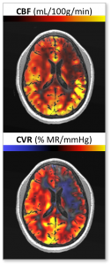
We use arterial spin labelling (ASL) MRI to non-invasively quantify regional cerebral blood flow (CBF) at the tissue level and generate high resolution 3D tissue perfusion maps. Furthermore, we assess the capacity for blood vessels to regulate regional blood flow. This is performed by measuring the relative change in CBF using blood-oxygen level dependent (BOLD) MRI in response to a global vasoactive stimulus such as CO2. The responsiveness of the vessels are expressed in terms of cerebrovascular reactivity (CVR) and can be mapped out for the entire brain.
The combination of CBF and CVR measurements provides important insight into the vascular physiology of the brain not only at baseline, but during physiological or metabolic stress. Current findings have shown that abnormal CBF and CVR are associated with brain injury and cognitive performance. As such, these are important factors for assessing vessel function in diseases affecting the cerebral vasculature such as sickle cell disease, obesity, obstructive sleep apnea, and moyamoya.
Blood-brain barrier integrity
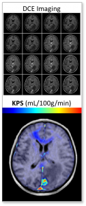
The blood-brain barrier (BBB) is an interface composed of endothelial cells connected by tight junctions that protect the brain from foreign substances in the bloodstream. Disruption of the BBB is indicative of cerebrovascular disease and can be a predictor of later complications. We use dynamic contrast-enhanced (DCE) MRI to map the integrity of the BBB throughout the brain. DCE-MRI is based on rapid sequential imaging of the circulation and accumulation of a contrast agent in the brain. The temporal profile of the contrast leakage into the brain can be used to quantitatively model the BBB integrity.
Studies have shown that the BBB is disrupted following ischemic stroke. Our lab has been able to quantify this damage in acute stroke patients and pre-clinical models of stroke using MRI combined with the appropriate pharmacokinetic model. We aim to elucidate the degree and progression of BBB disruption after stroke in the paediatric population, and investigate the efficacy of BBB stabilizing treatments during the acute phase of stroke.
Cerebral oxygen metabolism
The cerebral metabolic rate of oxygen (CMRO2) is an important physiological measure that quantifies the energy being used in the brain. The body maintains a consistent CMRO2 through a precise balance between cerebral blood flow, arterial blood oxygen content, and oxygen extract fraction (OEF). Recent advances in MRI have allowed us to non-invasively assess these parameters within clinically feasible scan durations. In particular, techniques for acquiring OEF, defined as the percent of oxygen in the blood that used by the brain, are continuously improving and is the subject of much research.
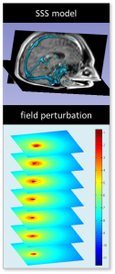
Our lab implements a novel technique that measures venous oxygen saturation using the MRI phase information in the superior sagittal sinus to compute global, and potentially regional, OEF. We are currently investigating CMRO2 changes in patients with sickle cell disease (SCD) and sleep disorders. Our study has shown SCD patients have reduced OEF compared to healthy controls, and distinct disease phenotypes may be at higher risk of future ischemic injury.
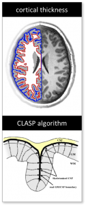
Cortical morphometry
Cortical thickness is a subtle but well established marker of brain development. Abnormal grey matter thickness has been associated with various diseases and may even correlate with cognitive function. However, the differences are difficult to detect in-vivo as the human cerebral cortex consists of multiple gyri and sulci with an average thickness of only a few millimetres.
High-resolution T1 weighted MRI can produce excellent contrast between the grey and white matter in the brain. Combined with advanced post-processing techniques (CIVET) and computational algorithms (CLASP), we can define the boundaries of the cortex and generate cortical surface maps of the brain. Our lab has demonstrated regional differences in cortical thickness between healthy children and children with sickle cell disease. We are currently exploring the effects of other diseases such as sleep disorders and stroke.

