Cellular and Molecular Electron Microscopy (CMEM)
The Cellular and Molecular Electron Microscopy (CMEM) core is a joint facility of SickKids Research Institute and the Lunenfeld-Tanenbaum Research Institute. The facility provides electron microscopy services to the scientific community, including research institutes and universities.
CMEM offers an extensive range of services for transmission and scanning electron microscopy (TEM and SEM) including sample processing for biological and organic specimen, negative stain imaging, consultation and student training.
Equipment
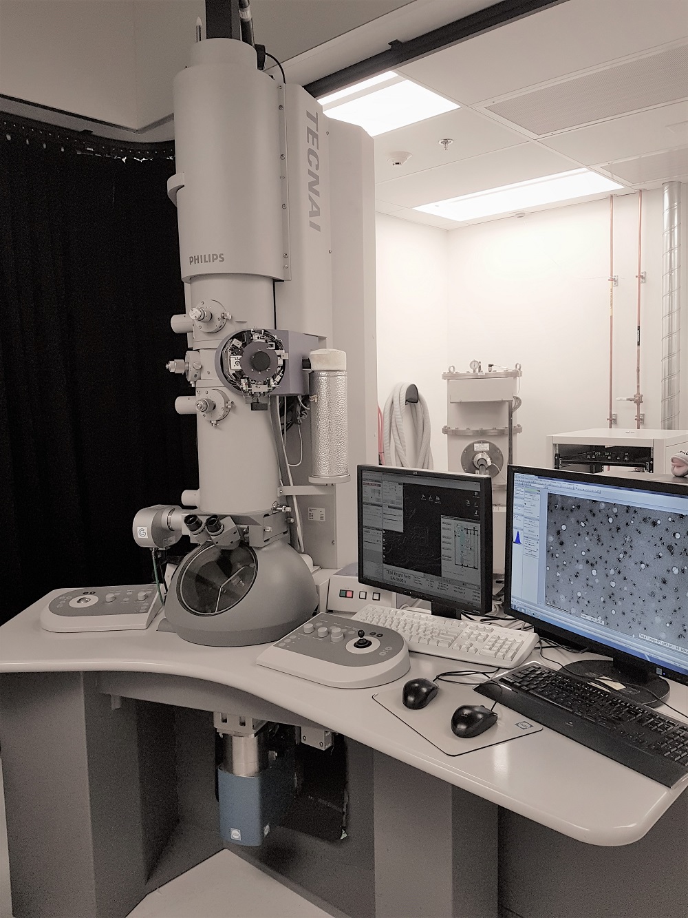
This is a 20kV scanning electron microscope with variable voltage capable of 4nm resolution at high vacuum. Low pressure operation is also available. The unit is equipped with secondary electron and back-scattered electron detectors. A user friendly machine to screen the topology and surface morphology of your specimen. The system features automatic beam alignment including auto focus and auto stigmation. Chamber view camera allows for imaging of 6 samples at a time.
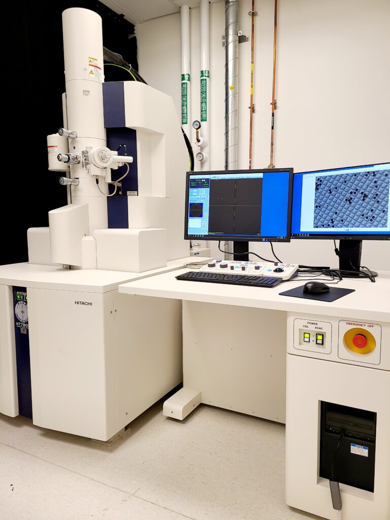 Hitachi HT7800 TEM is the revolution of ultimate luxury TEM imaging. This 120 kV microscope with dual objective lens enables both High Resolution and High Contrast imaging. Advanced stage-navigation function provides whole-grid searching and efficient image acquisition.
Additional functions include Automated Image Stitching, 3D Electron Tomography, Drift correction, Panoramic imaging and with the use of multi specimen holders users can load up to 3 grids each run.
This microscope is paired with the EMSIS Xarosa 20 Megapixel CMOS camera which provides high dynamic range imaging at 30 FPS.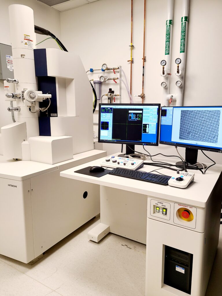
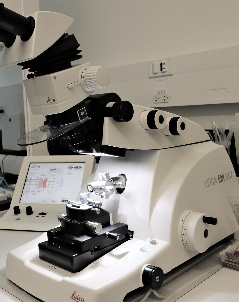 The Leica UC7 is capable of cutting thin sections on resin embedded cells and tissues from 50nm to semi-thin sections of few micron. We will use wet diamond and ionizer to ensure clean and artefact free sections.
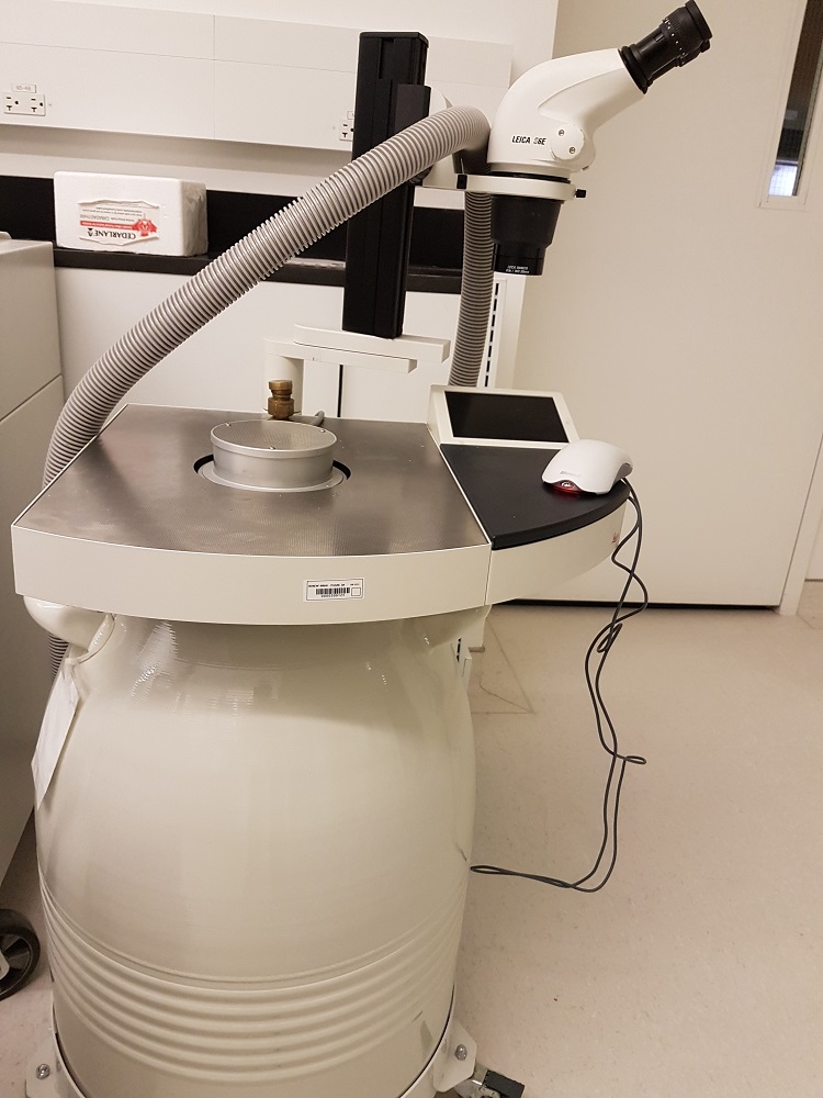
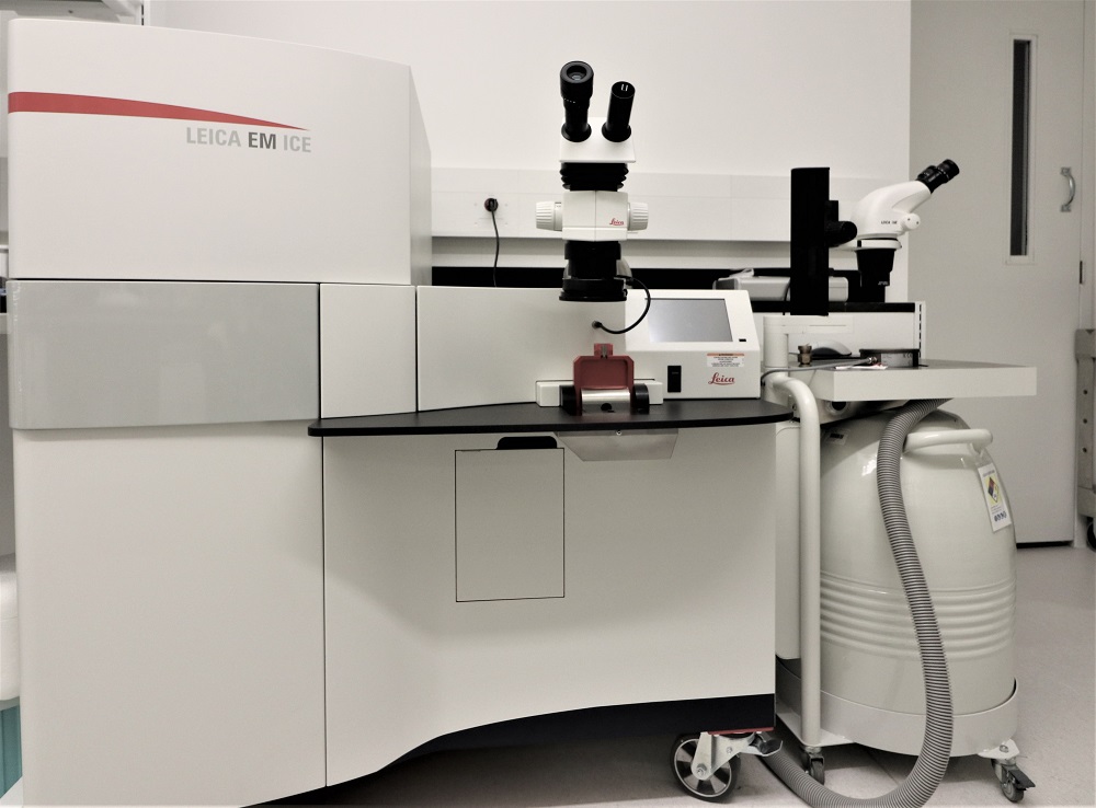 High pressure freezing technique is used to vitrify cells and specimen below 200um thickness. This method employs a fast freezing to liquid nitrogen temperature at high pressure of 2100 bar to insure near life like preservation of ultrastructure. Copper or Sapphire carriers of 3 or 6 mm diameters are used for various applications including correlative election light microscopy (CLEM).
 Following high pressure freezing the specimen is processed via freeze substitution where the frozen water is replaced by fixative and post stain reagents at cryo temperature and controlled speed. This machine is equipped with UV lamp for resin curing at cryo or room temperature.
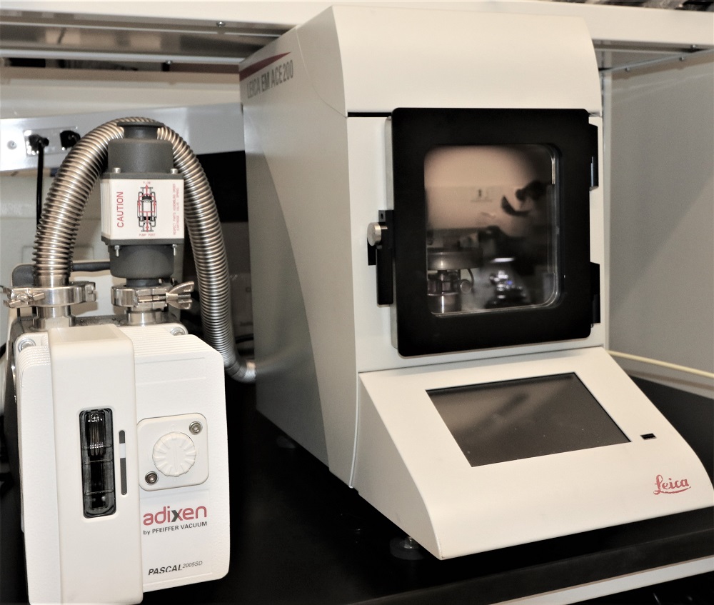 Gold sputtering on specimen at precisely controlled rate using diffusion or direction method.
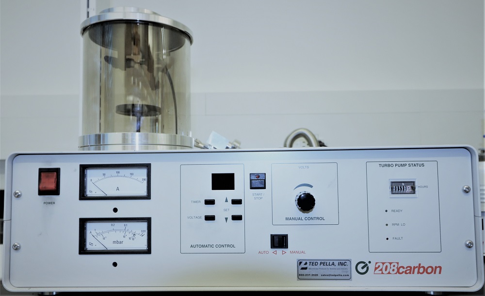 Pulsed deposition of carbon film on specimen at high vacuum.
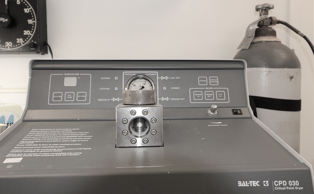 CPD method is used to dry samples prepared for SEM imaging. The machine will dry the dehydrated specimen using liquid carbon dioxide at 70 PSI and 42 degrees Celsius.
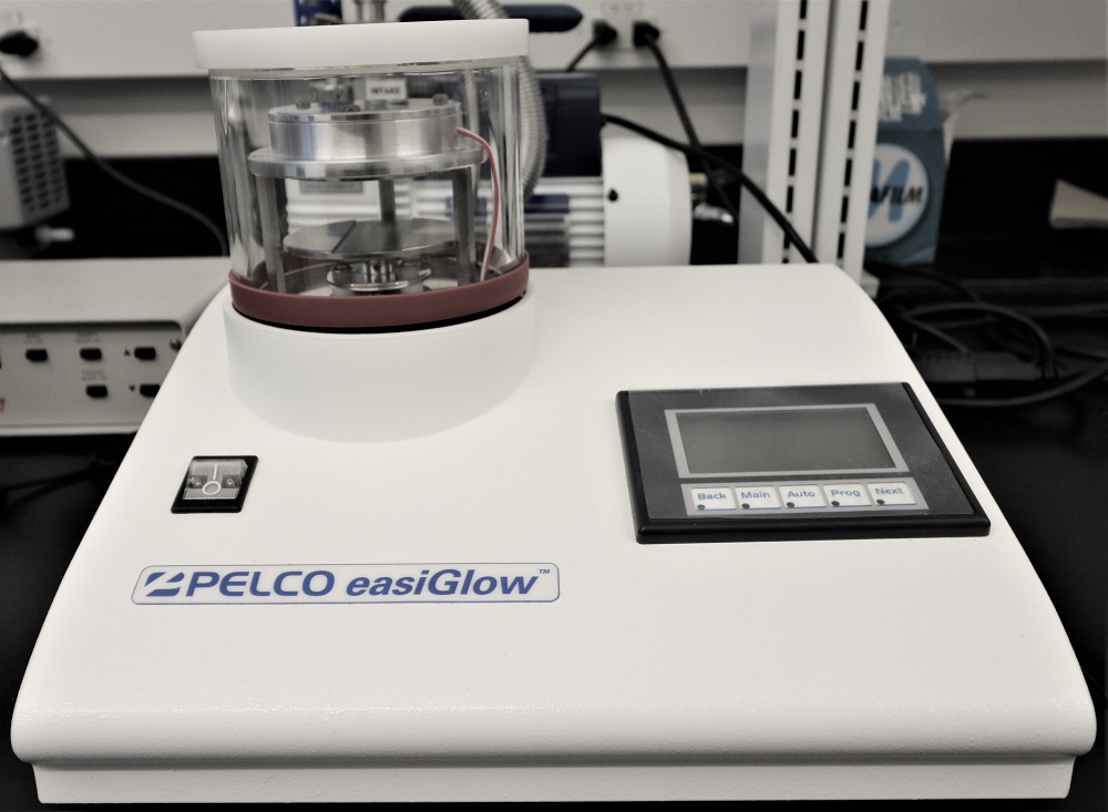 Plasma cleaning of TEM grids or SEM specimen. The tool is capable of both positive and negative discharge.
Fees
| Service/equipment | Price |
|---|---|
| Technologist’s time | $60/hr |
| Tecnai TEM | $60/hr |
| Phoenix TEM | $60/hr |
| Hedwig TEM | $60/hr |
| Preparation equipment | Price |
|---|---|
| Ultramicrotome | $50/hr |
| High-pressure freezer | $250/10 shots |
| Freeze substitution | $50/day |
| Sputter coater | $22/run |
| Carbon evaporator | $22/run |
| Critical point dryer | $22/run |
| Specimen preparation | Price |
|---|---|
| TEM preparation | $65/specimen |
| SEM preparation (fixation, critical point drying, sputter coat, immunostaining) |
$65/specimen |
| Semi-thin sections | $13/block |
| Thin sections | $27/block |
| ** Industrial rate is cost X2 |
Contact and bookings
For same-day preparation and training, please contact Dr. Ali Darbandi, Facility Manager at ali.darbandi@sickkids.ca
Facility Address: 686 Bay St, Peter Gilgan Centre for Research and Learning, 6th floor, Room 9621. Toronto, ON, Canada M5G 0A4
All equipment reservation is done through QReserve, our online booking calendar.
Standard operating procedures (SOPs)
Team


Dr. Mei Zhen
zhen@lunenfeld.ca
416-586-4800

Dr. Ali Darbandi, Facility Manager
ali.darbandi@sickkids.ca
416-813-7654 ext. 309970
Recent publications
- K. Battiston et al, Nature communications, 12, 2875 (2021)
- M. Mahendralingam et al, Nature Metabolism, 3, 665-681 (2021)
- L. Antounians et al, Science Translational Medicine, 13, issue 550 (2021)
- M. Ebrahimi et al, Acta Biomaterilia, 132, 227-244 (2021)
- L. Ermini et al, Front. Cell. Dev. Biol. 91 652651 (2021)
- T. Li et al, Fertility and Sterility, 115, 1327-1336 (2021)
- A. Karunendiran et al, BMC Molecular and Cell Biology, 22:38 (2021)
- D. Fernandes et al, RSC Adv, 11, 4906-4920 (2021)
- J. Xu et al, Small, 17, 2100345 (2021)
- C. Daeschler et al, CMAJ, 192, 42 (2020)
- Ngo et al, J. Am. Chem. Soc 142, 42, 17938–17943 (2020)
- Zhang et al, CS Nano, 14, 8, 9478–9490 (2020)
- Ouyang et al, Nature Materials, The dose threshold for nanoparticle tumour delivery (2020)
- Smith et al, PLOS ONE, 15(8), e0231364 (2020)
- Soeandy et al, Cellular and Molecular Neurobiology,
Necroptotic–Apoptotic Regulation in an Endothelin-1 Model of Cerebral Ischemia (2020)
- Rao-Bhatia et al, Developmental Cell, 52,5, 647-658 (2020)
- Chung et al, Nature Communications, 11, 825 (2020)
- Antounians et al, BioRxiv, Impaired Fetal Lung Development can be Rescued by Administration of Extracellular Vesicles Derived from Amniotic Fluid Stem Cells (2020)
- Balakrishnan et al, BioRxiv, SMPD3-mediated extracellular vesicle biogenesis inhibits oligodendroglioma growth (2020)
- Steadman et al, Neuron, 105,1, 150-164 (2020)
- Fievel, University of Toronto Thesis, (2020)
- Ghomashchi, University of Toronto Thesis, (2020)
- Mulcahy et al, Neural Circuit 13,16 (2019)
- Poon et al, ACS Nano, 13, 5, 5785-5798 (2019)
- Fernandes et al, Optical Materials Express, 9,12, 4532-4544 (2019)
- Vijayakumar et al, Nanoscale Horiz, 4, 495-515 (2019)
- Lazarovits et al, ACS Nano, 13, 7, 8023-8034 (2019)
- Sandoval et al, Int J Clin Exp PAthol 12, 5, 1713-1722 (2019)
- Coindre et al, Advanced Healthcare Materials, 8, 18 (2019)
- Overchuk et al, ACS Nano, 13,4,4560-4571 (2019)
- Reunov et al, Differentiation, 109, 34-41 (2019)
- Condori et al, eLife 8, e47918 (2019)
- Coquenlorge et al, Cell Reports, 27,10, 3006-3018 (2019)
- Kolosov, et al, Journal of Experimental Biology, 222 (2019)
- Zhao et al, Human Molecular Genetics, 28, 24 4186-4196 (2019)