3D Scanning Electron Microscopy (3D SEM)
The 3D SEM core provides cutting-edge scanning electron microscopy (SEM) equipment. We employ the latest generation of electron microscopes with advanced technology to support revolutionary cell biology research. We offer fast image acquisition, high resolving power and the ability to generate 3-dimensional images in a world-class laboratory.
Equipment
Zeiss Gemini 2 SEM
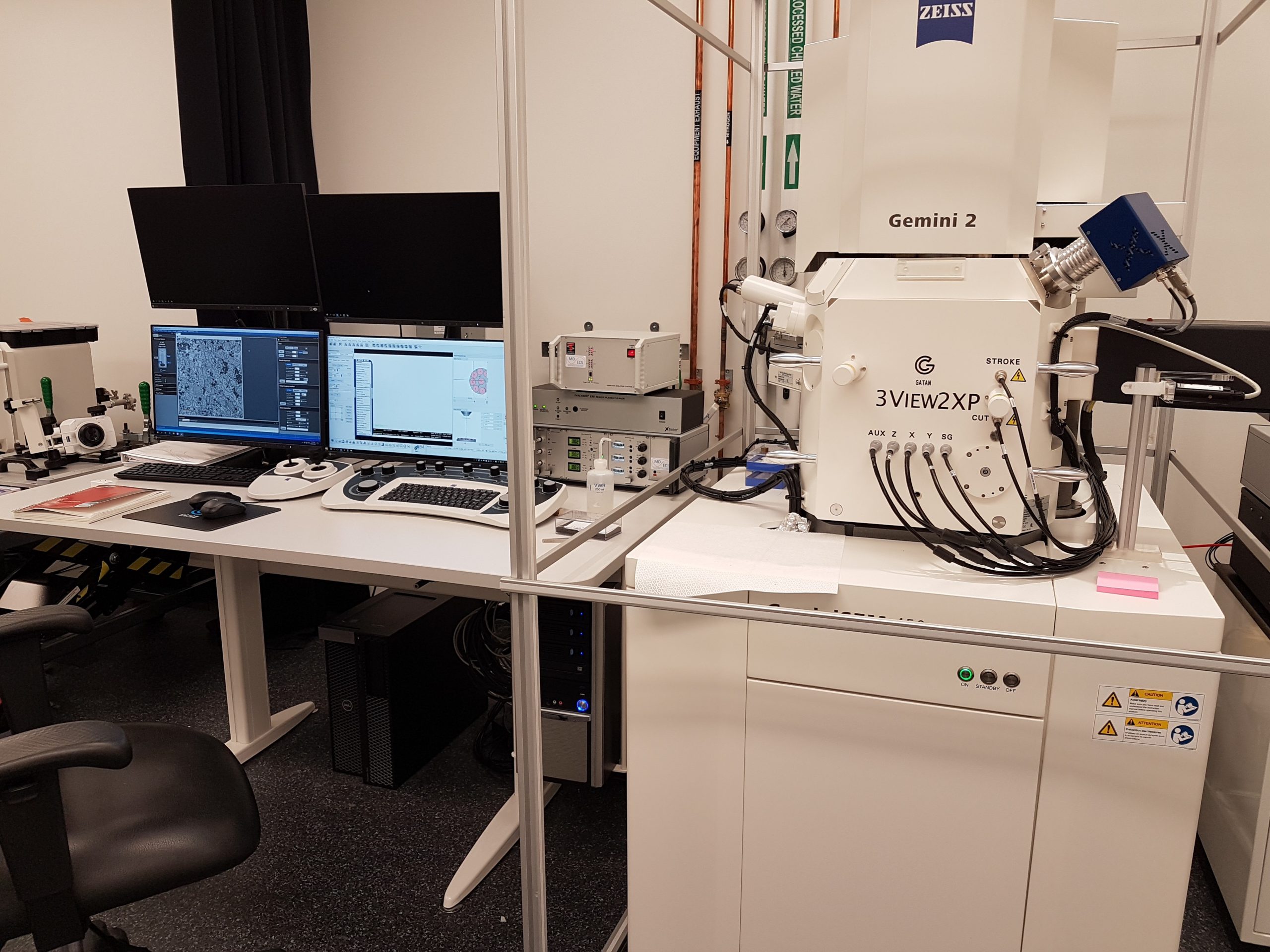
The Gemini is a field emission gun microscope equipped with the Gatan 3View serial block face for Automate sectioning and image capture of 3D ultrastructures.
It is capable of imaging at low voltages below 1kV with very small probe sizes and high signal-to-noise (SNR) ratio thanks to novel beam booster technology.
There are five detectors available on this machine:
- Secondary electron detector
- Inlens SE detector
- Backscattered electron detector, four-segment annular design
- Inlens Energy selective backscattered electron detector
- OnPoint Gatan backscattered electron detector for block face imaging
Zeiss Crossbeam 550
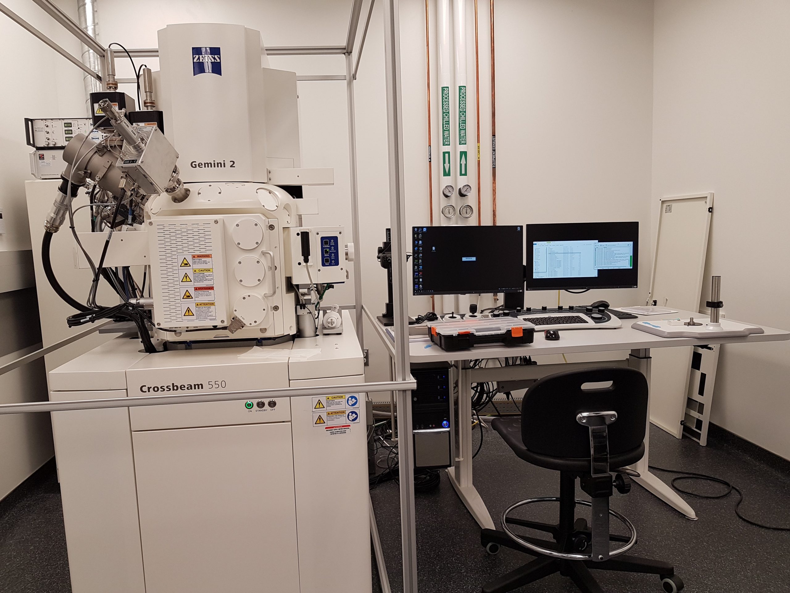
The Crossbeam is a field emission scanning electron microscope with the processing ability of a focused ion beam.
This instrument uses a Gallium focused ion beam for serial milling of samples and sequential SEM imaging. This beam can remove sections as thin as 5 nm, providing the highest possible z-plane resolution of biological samples.
There are six key features available on this machine:
- SESI detector for secondary electron and secondary ion imaging
- Inlens SE detector
- Backscattered electron detector, four-channel parallel annular design
- Inlens Energy selective backscattered electron detector
- Two channels Gas Injection System (GIS) for Platinum and Carbon deposition
- Annular STEM detector for transmission imaging
Services
Serial Block Face (SBF) imaging
3D SBF imaging involves the use of an ultramicrotome mounted inside the vacuum chamber of a SEM to cut a thin section from the face of the block. After the section is cut, the specimen is raised back to the focal plane and imaged again. This sequence of specimen imaging, section cutting and block raising can acquire thousands of images in perfect alignment in an automated fashion.
Array Tomography and Correlative Light Electron Microscopy
It can be challenging to determine where specific molecules, like proteins or lipids, are located relative to cell structures. Correlative light-EM (CLEM) imaging is one method used to do this. CLEM merges the power of light microscopy (with its many fluorescentreporters of protein/lipid location) with the resolving power of EM. The ATLAS 5 software includes dedicated software for semi-automated SEM imaging of serial sections at multiple resolutions for Array Tomography. ATLAS 5 software enables advanced, correlative microscopy by fusion of data sets from multiple instruments, detectors and sessions into one correlative workspace.
FIB-SEM Imaging
Our facility can image at high-resolutions while milling with a focused ion beam. The Atlas 5 3-D nanotomography package allows for simultaneous milling and imaging without having to switch off either beam during the entire acquisition process. The resulting high z-resolution can visualize membrane alterations and their association with other cellular structures. We can employ high pressure freezing to provide ideal sample preservation and minimize potential imaging artefacts.
Fees
The 3D SEM core provides high quality service, competitive prices, and a wide range of available techniques. We offer consultation, sample preparation, user training and data acquisition.
Note: The regular rates are reduced to a flat $220 /day for academic users after 10 days of usage. The usage can be non-consecutive.
| Service | Internal cost | External cost | Industry cost |
|---|---|---|---|
| Gemini SEM | $640/day | $780/day | $1,280/day |
| Crossbeam SEM | $640/day | $780/day | $1,280/day |
| Technologist’s time | $70/hour | $70/hour | $140/hour |
Contact and bookings
In order to access the 3D SEM services, new clients must submit an application form to describe their projects and approach. The facility manager and directors will consult the client on suitable imaging methods and sample preparation.
Once the application has been approved, samples will be processed in the CMEM sub-core and screened for quality using our TEM.
Successful samples will then be prepared for volume imaging and imaged at the 3D SEM facility.
For questions or more information about our services, please contact Dr. Ali Darbandi at ali.darbandi@sickkids.ca
Team


Dr. Ali Darbandi, Facility Manager
ali.darbandi@sickkids.ca
416-813-7654 ext. 309970